System and method for image based physiological monitoring of cardiovascular function

a physiological monitoring and image-based technology, applied in image analysis, medical science, image enhancement, etc., can solve the problems of non-diagnostic ischemia detection, non-reporting of ischemic changes on electrocardiogram (ecg) during dobutamine stress testing or mr-guided intervention, and inability to detect ischemia, etc., to achieve more sensitive feedback and increase sensitivity

Inactive Publication Date: 2013-11-05
SIEMENS MEDICAL SOLUTIONS USA INC
View PDF12 Cites 4 Cited by
  • Summary
  • Abstract
  • Description
  • Claims
  • Application Information

AI Technical Summary

Benefits of technology

[0006]Exemplary embodiments of the invention as described herein generally include methods and systems for real-time assessment of cardiac function by 2D MR imaging. Cardiac function can be assessed visually, or displayed as m-mode data to enable assessment of the temporal history as well. The m-mode display and resulting measurements can be interleaved with other types of acquisition and incorporated into a real time interface for scanner control. A system and method of an embodiment of the invention enable early ischemia detection based on wall motion, using real time imaging interleaved with ‘standard’ imaging. By interleaving MR data acquisition one can use a portion of the data for direct heart function monitoring, on a continuous basis, while the rest of the data are used for diagnostic or interventional purposes. Wall motion and ventricular function parameters are extracted from the real time imaging, and methods for change detection alert the operator that heart function has changed. In addition to monitoring for ischemia, methods according to embodiments of the invention can be used to detect changes in patterns of contraction (due to conduction abnormalities), detect changes in cardiac output (real time flow imaging), or changes in patterns of perfusion (real time myocardial perfusion imaging). Embodiments of the invention extract relevant features from the interleaved monitoring data, and identify clinically relevant changes to assist the operator in monitoring heart function. A system according to an embodiment of the invention for real-time image interleaving can provide near-continuous monitoring of heart function in the MR environment where ECG is not diagnostic. In addition, it can potentially provide more sensitive feedback on changes in heart function than global ECG monitoring could provide, independent of its use in an MRI environment. This potentially increased sensitivity stems from the fact that both wall motion changes and myocardial perfusion changes precede ECG changes during ischemia. The concept of using direct measurement of heart function in the background while some other diagnostic procedure or therapeutic imaging is being performed can also be applied to other imaging modalities, such as computed tomography (CT) or ultrasound (US).
[0007]Validation results show the feasibility of a real time method for global left ventricle function assessment that can be combined with stress testing or interventional procedures. Integration into a real time environment using 1-2 slices for function monitoring and additional temporally interleaved slices for procedure guidance is feasible.

Problems solved by technology

Monitoring the electrocardiogram (ECG) for ischemic changes during dobutamine stress testing or an MR-guided intervention is not possible in the MR environment due to the magnetohydrodynamic effect.
This additional voltage is added to the ECG when the patient is inside the static magnetic field, causing distortion.
The main challenge in patient monitoring for ischemia in MRI is that the static magnetic field, RF pulses, and field gradient switching all distort the ECG making it non-diagnostic for ischemia detection.
Ischemia in the heart first results in a perfusion deficit, then in abnormalities in wall motion, then ECG changes, then finally in chest pain.
During interventional procedures, changing the real time scan plane is used to check for changes in function, but this does not provide continuous monitoring.
Visual review of perfusion scans during acquisition also can be used to check for changes in heart perfusion, but these cannot be repeated as often during an exam as ventricular function imaging since they require contrast agent administration.

Method used


Image

  • System and method for image based physiological monitoring of cardiovascular function
  • System and method for image based physiological monitoring of cardiovascular function
  • System and method for image based physiological monitoring of cardiovascular function

Examples


Embodiment Construction

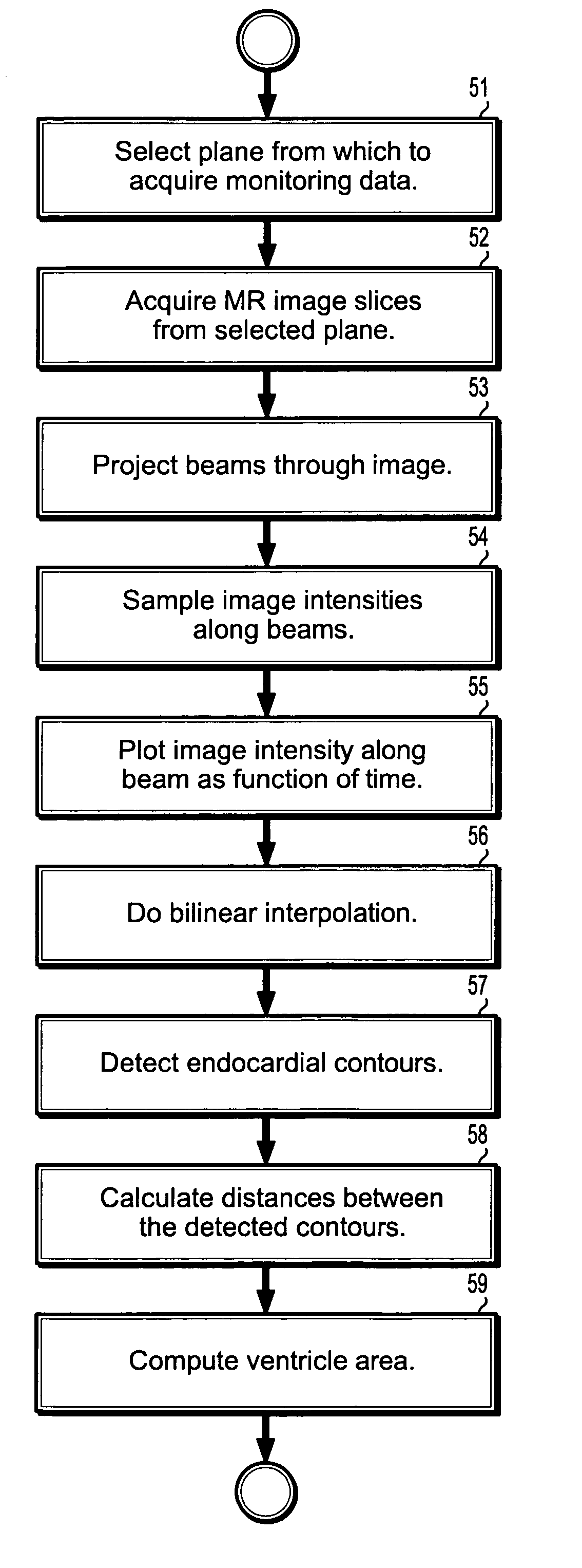
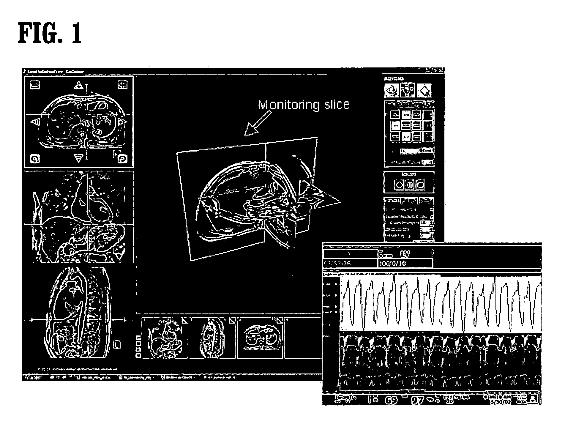
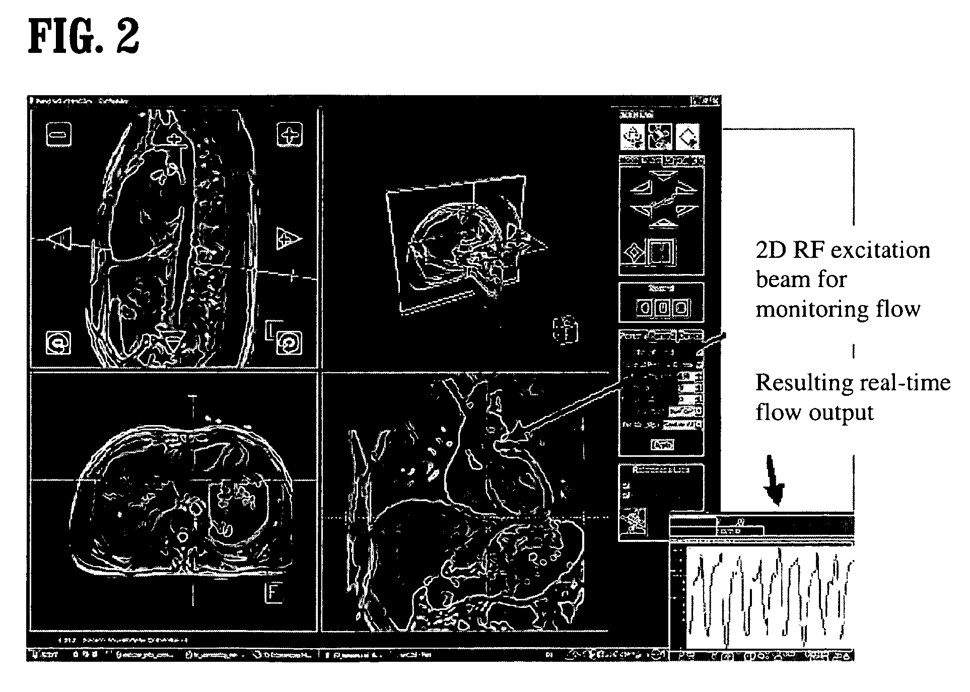
[0015]Exemplary embodiments of the invention as described herein generally include systems and methods for real-time monitoring of heart function using magnetic resonance imaging (MRI), interleaved with imaging used to guide or perform stress testing or interventional cardiovascular procedures. Accordingly, while the invention is susceptible to various modifications and alternative forms, specific embodiments thereof are shown by way of example in the drawings and will herein be described in detail. It should be understood, however, that there is no intent to limit the invention to the particular forms disclosed, but on the contrary, the invention is to cover all modifications, equivalents, and alternatives falling within the spirit and scope of the invention.

[0016]As used herein, the term “image” refers to multi-dimensional data composed of discrete image elements (e.g., pixels for 2-D images and voxels for 3-D images). The image may be, for example, a medical image of a subject co...


PUM


Abstract

A method for real time monitoring of cardiac function includes defining a plurality of planes through a heart from which to acquire heart imaging data, acquiring a sequence of 2-dimensional images from said selected planes, wherein each said image comprises a plurality of intensities defined on a domain of points on a 2D grid, selecting one or more of said sequence of images to provide imaging data for monitoring heart function, wherein said remaining unselected images are adapted for diagnostic purposes, and repeating said steps of acquiring a sequence of 2-dimensional images from said selected planes and selecting one or more of said sequence of images for monitoring heart function, wherein a time sequence of images is obtained, alternating between images for monitoring heart function and images for diagnostic purposes.

Description

CROSS REFERENCE TO RELATED UNITED STATES APPLICATIONS[0001]This application claims priority from: “Image Based Physiological Monitoring of Cardiovascular Function”, U.S. Provisional Application No. 60 / 749,558 of Lorenz, et al., filed Dec. 12, 2005, the contents of which are incorporated herein by reference.TECHNICAL FIELD[0002]This invention is directed to real-time monitoring of heart function using magnetic resonance imaging (MRI), interleaved with imaging used to guide or perform stress testing or interventional cardiovascular procedures.DISCUSSION OF THE RELATED ART[0003]Monitoring the electrocardiogram (ECG) for ischemic changes during dobutamine stress testing or an MR-guided intervention is not possible in the MR environment due to the magnetohydrodynamic effect. This effect occurs because ions in the blood flowing through the body in the presence of a static magnetic field create its own electrical voltage. This additional voltage is added to the ECG when the patient is insi...

Claims


Application Information

Patent Timeline
Patent Type & Authority Patents(United States)
IPC IPC(8): A61B5/05
CPCA61B5/055G06T7/0083G06T7/0089G06T7/20G06T2207/10088G06T2207/30048G06T7/12G06T7/149
Inventor LORENZ, CHRISTINE HMAIER, CORINNA
Owner SIEMENS MEDICAL SOLUTIONS USA INC
Why Eureka
Try Eureka