System and method for segmenting bones on MR images

a technology of magnetic resonance imaging and bone segmentation, applied in image enhancement, image analysis, instruments, etc., can solve the problems of false classification of tissue and organs, difficult task of separating bone structures from other tissues on mr images, and time-consuming and laborious combining to form 3d volumes, etc., to achieve improved performance and reliability.

Active Publication Date: 2013-11-19
GENERAL ELECTRIC CO
View PDF7 Cites 12 Cited by
  • Summary
  • Abstract
  • Description
  • Claims
  • Application Information

AI Technical Summary

Problems solved by technology

Manually drawing the individual contours on a contiguous set of 2D slices and combining them to form 3D volumes is very time consuming and labor intensive.
However, if MR images are used, it is a difficult task to separate bone structures from other tissues on MR images.
Many existing segmentation techniques for extracting bones on MR images, however, have disadvantages such as a risk of falsely classifying tissue and organs as bone and are not able to be automated.

Method used


Image

  • System and method for segmenting bones on MR images
  • System and method for segmenting bones on MR images
  • System and method for segmenting bones on MR images

Examples


Embodiment Construction

[0014]In the following detailed description, numerous specific details are set forth in order to provide a thorough understanding of embodiments. However it will be understood by those of ordinary skill in the art that the embodiments may be practiced without these specific details. In other instances, well-known methods, procedures, components and circuits have not been described in detail so as not to obscure the embodiments.

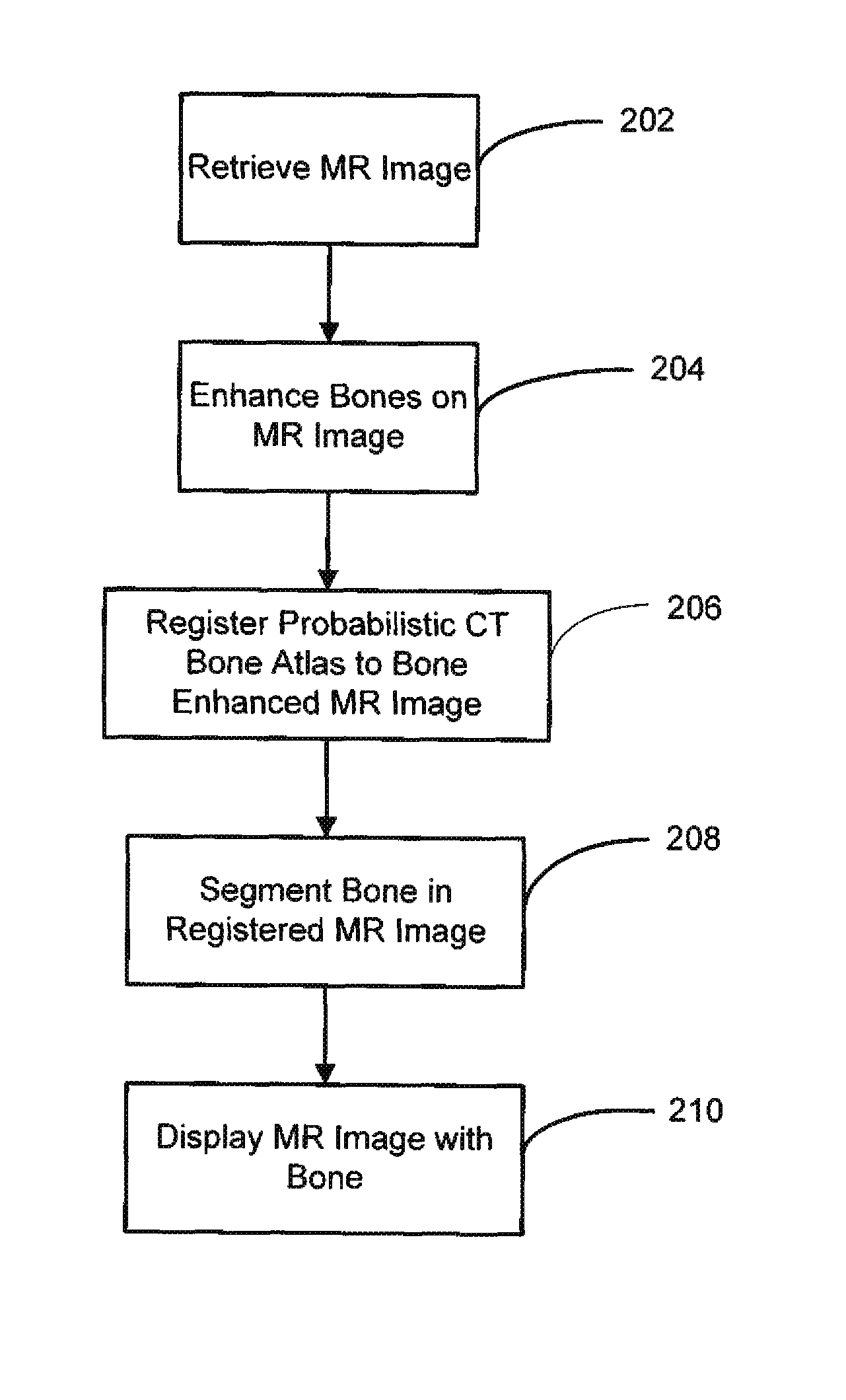
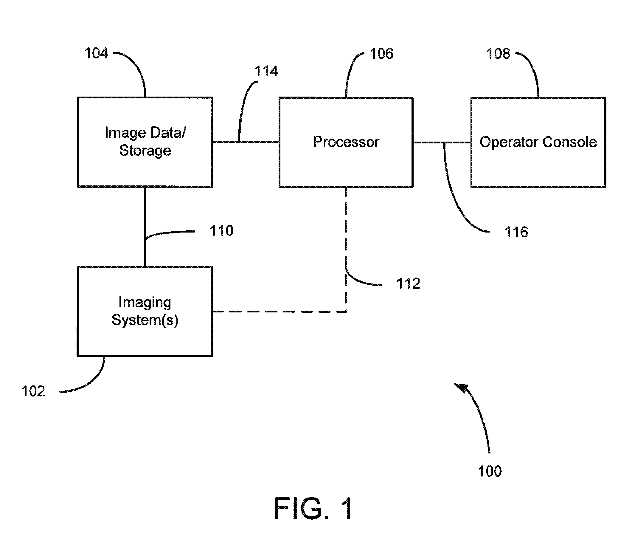
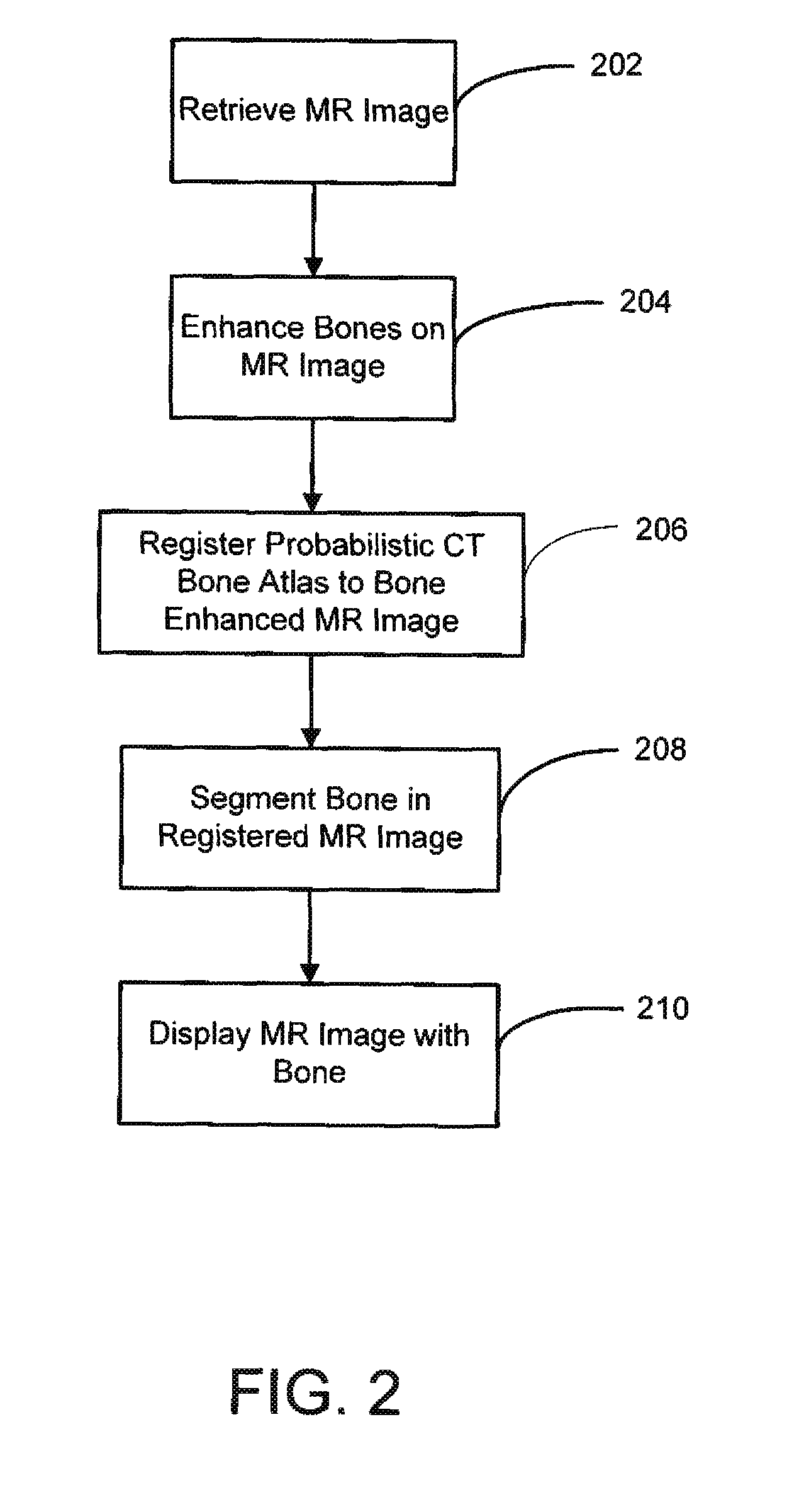
[0015]FIG. 1 is a schematic block diagram of a system for automatic segmentation of bone on MR images in accordance with an embodiment. In system 100, image data is stored in storage or memory 104. Storage 104 may be capable of storing sets of data or images generated by one or more imaging systems 102 or images and data generated using processor 106 and operator console 108. Storage 104 may be, for example, integrated into the imaging system 102 or processor 106 or may be remotely located and connected to the imaging system 102 and processor 106 through a net...


PUM


Abstract

A method for segmenting bones on magnetic resonance (MR) images includes retrieving an MR image and performing an enhancement process on the MR image to generate a bone enhanced MR image. The bone enhanced MR image is then registered to a computer tomography (CT) based bone atlas. An MR image with bone segmentation is generated by segmenting the bone enhanced MR image using the CT based bone atlas as a mask. The MR image with bone segmentation may be presented on a display.

Description

CROSS REFERENCE TO RELATED APPLICATION[0001]The present application claims the benefit of U.S. Provisional Application Ser. No. 61 / 541,801, filed Sep. 30, 2011, herein incorporated by reference in its entiretyTECHNICAL FIELD[0002]The present invention relates generally to magnetic resonance imaging (MRI) systems and in particular, to a system and method for automatically segmenting bones on MR images.BACKGROUND[0003]During clinical diagnosis, the internal anatomy of a patient is imaged to determine how a certain disease has progressed. For example, the images may be used to help distinguish between infected tissues and healthy tissues within the patient. The images are also useful for radiotherapy treatment or planning or for surgical planning. Several modalities are used to generate images or functionality of anatomy of a patient which are suitable for diagnostic purposes, radiotherapy treatment or surgical planning. Examples include conventional X-ray imaging, computed tomography ...

Claims


Application Information

Patent Timeline
Patent Type & Authority Patents(United States)
IPC IPC(8): G06K9/00
CPCG06T7/0083G06T7/0087G06T7/0097G06T2207/10081G06T2207/10088G06T2207/20076G06T2207/20128G06T2207/30008G06T7/12G06T7/143G06T7/174
Inventor NOVAK, GABORREDELE, MILANFIDRICH, MARTAUNGI, TAMAS
Owner GENERAL ELECTRIC CO
Why Eureka
Try Eureka