

SAR dosimeter for RF power deposition in MRI and methods and systems related thereto
a technology of rf power deposition and dosimeter, which is applied in the field of systems, devices and methods used in magnetic resonance imaging, can solve the problems of incorrect scanner sar, increased risk, and inability to accurately measure the true average of scanner sar, etc., and achieves the effect of less costly use and simple construction
- Summary
- Abstract
- Description
- Claims
- Application Information
AI Technical Summary
Benefits of technology
Problems solved by technology
Method used
Image

Examples
example 1
Methods
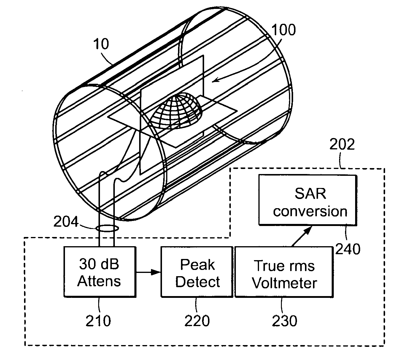
[0087]Transducer:
[0088]A prototype 1.5 T SAR dosimeter transducer that is accommodated in the bore of a head coil for measuring average head SAR, is fabricated from two 18-cm square copper loops, affixed to acrylic board. The loops are oriented orthogonal to each other to permit detection of both components of a quadrature excitation field in the XY-plane, should one exist. They are tuned to the 1.5 T MRI frequency, and loaded with resistors. The center of each board is removed to accommodate a small spherical CuSO4-doped water phantom whose only purpose is to generate just enough MRI signal for the scanner to adjust its pulse power level and / or set-up the pulse sequence flip-angles. The transducer formed by each loop is such as that illustrated in FIG. 2A and the circuit diagram of the loop is shown in FIG. 4.
[0089]To determine the value of the total load resistance, RL, for each loop such that the transducer presents a load equivalent to that of the head to the MRI scanner,...
PUM
 Abstract
Description
Claims
Application Information