Radiation dosimetry apparatus and method, and dosimeter for use therein

a technology of radiation dosimetry and dosimeter, which is applied in the field of radiation dosimetry apparatus and methods for measuring radiation, and to dosimeters, and can solve the problems of reducing the accuracy and precision of radiation delivery, affecting the accuracy of radiation delivery, and affecting the accuracy of dose delivery and position accuracy

Active Publication Date: 2009-01-08
BEST THERATRONICS
View PDF10 Cites 53 Cited by
  • Summary
  • Abstract
  • Description
  • Claims
  • Application Information

AI Technical Summary

Benefits of technology

[0025]Thus, dosimetry apparatus and a method are provided employing a dosimeter having at least one radiation detector and at least one magnetic position sensor located a predetermined distance apart, so that radiation level readings from the radiation detector and position readings from the position sensor may be monitored and correlated, conveniently according to time, to obtain the position of the radiation detector when a particular radiation level was detected.

Problems solved by technology

The precision of delivery is affected during treatment by the inherent limitation of the imaging procedure used to delineate tumour boundary and the physician's skill in defining gross tumour volume; the latter varying from one physician to another.
The dose delivery accuracy and position accuracy may also be affected by the accuracy of the dose calculation algorithm, the daily patient setup errors and mechanical positioning tolerances of the treatment machine.
These effects individually or combined, will contribute to reduced accuracy and precision in the delivery of the radiation.
Further inaccuracy may be caused by movement of the target, i.e., the tumour, before and / or during therapy.
Although feasible, it would be undesirable for the physician to compensate by increasing the irradiated volume to ensure that the entire tumour is treated, because that would likely result in increased damage to neighbouring healthy tissue.
According to United States published patent application number 2006 / 0093089, the entire contents of both of which are incorporated herein by reference, imaging techniques, such as X-ray, CT, MRI and ultrasound, which are used to align the target volume with fiducial marks upon the patient and with the isocentre of the radiation beam equipment, are not particularly suitable for monitoring movement of the target volume caused by bodily functions.
It is not enough, however, merely to ensure that the radiation is applied to the right location.
The radiation level measured by a particular dosimeter detector may be affected by its proximity to an interface between two heterogeneous media and / or its proximity to a large dose gradient where dose measurements are difficult to perform accurately.
Even a slight body movement might result in the boundary of the prostate gland shifting and the rectum or urethra being subjected to an unacceptable level of radiation.
Nevertheless, the procedure is quite complex and provides the position at only one point in time.
The above-mentioned imaging techniques are not generally suitable for monitoring the location of a radiation detector during a radiation session.
More particularly, to monitor the position of the radiation detectors while taking account of organ movement would require repeated or even continuous imaging, since a single image would not be sufficient to correlate the organ movement and the position of the detector(s).
Also, it is not desirable to expose the patient to ionizing radiation in addition to that to which the patient must be exposed during therapy.
Nor is it usually convenient to use an X-ray machine or other imaging machine at the same time as the radiation therapy machine.
Changes also may occur during the actual implantation procedure as a result of edema and variable prostate texture causing movement in response to needle insertion and seed migration after insertion.
As before, real-time imaging to track movement of the radiation detector(s) is not practical.

Method used


Image

  • Radiation dosimetry apparatus and method, and dosimeter for use therein
  • Radiation dosimetry apparatus and method, and dosimeter for use therein
  • Radiation dosimetry apparatus and method, and dosimeter for use therein

Examples


Embodiment Construction

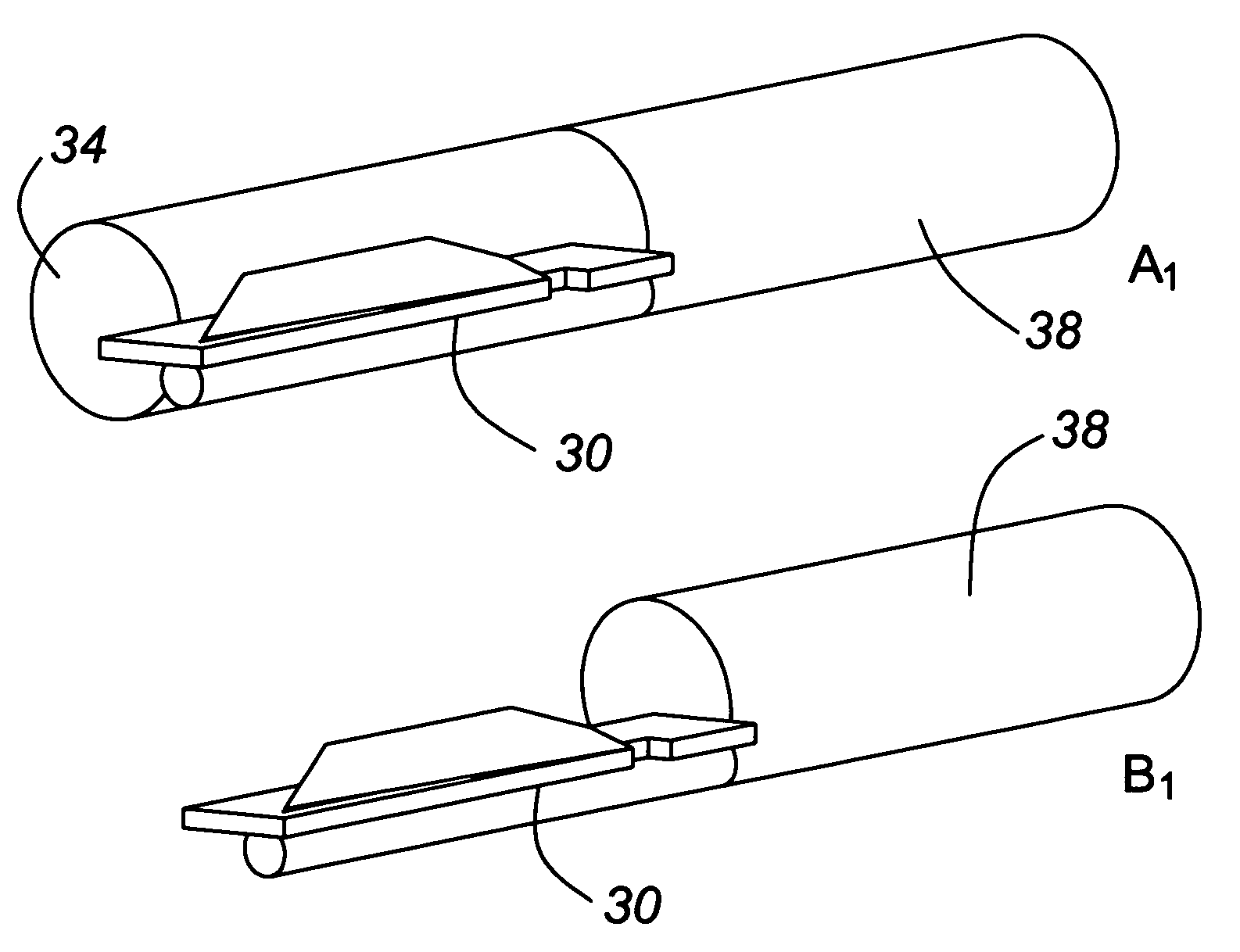
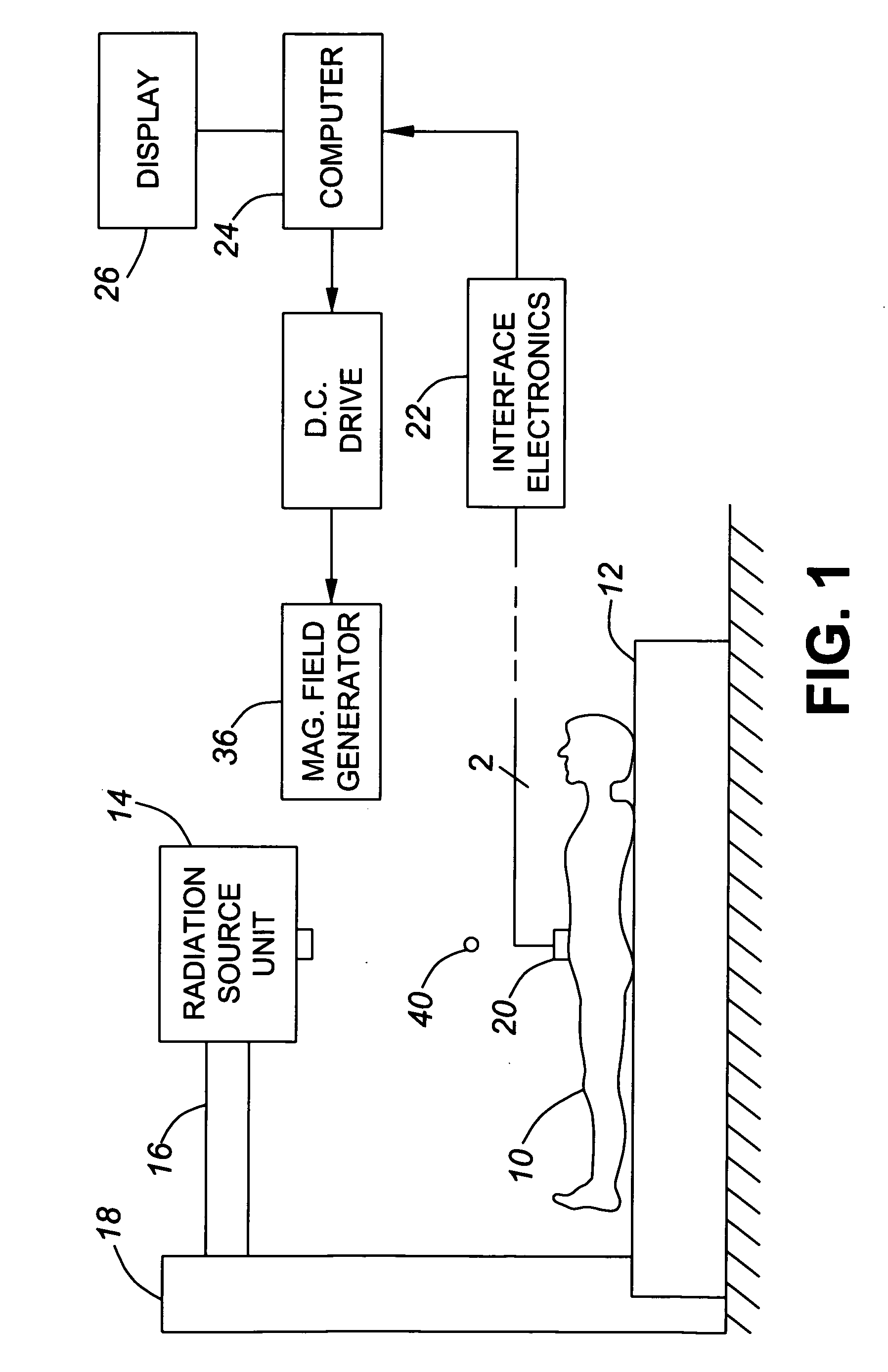
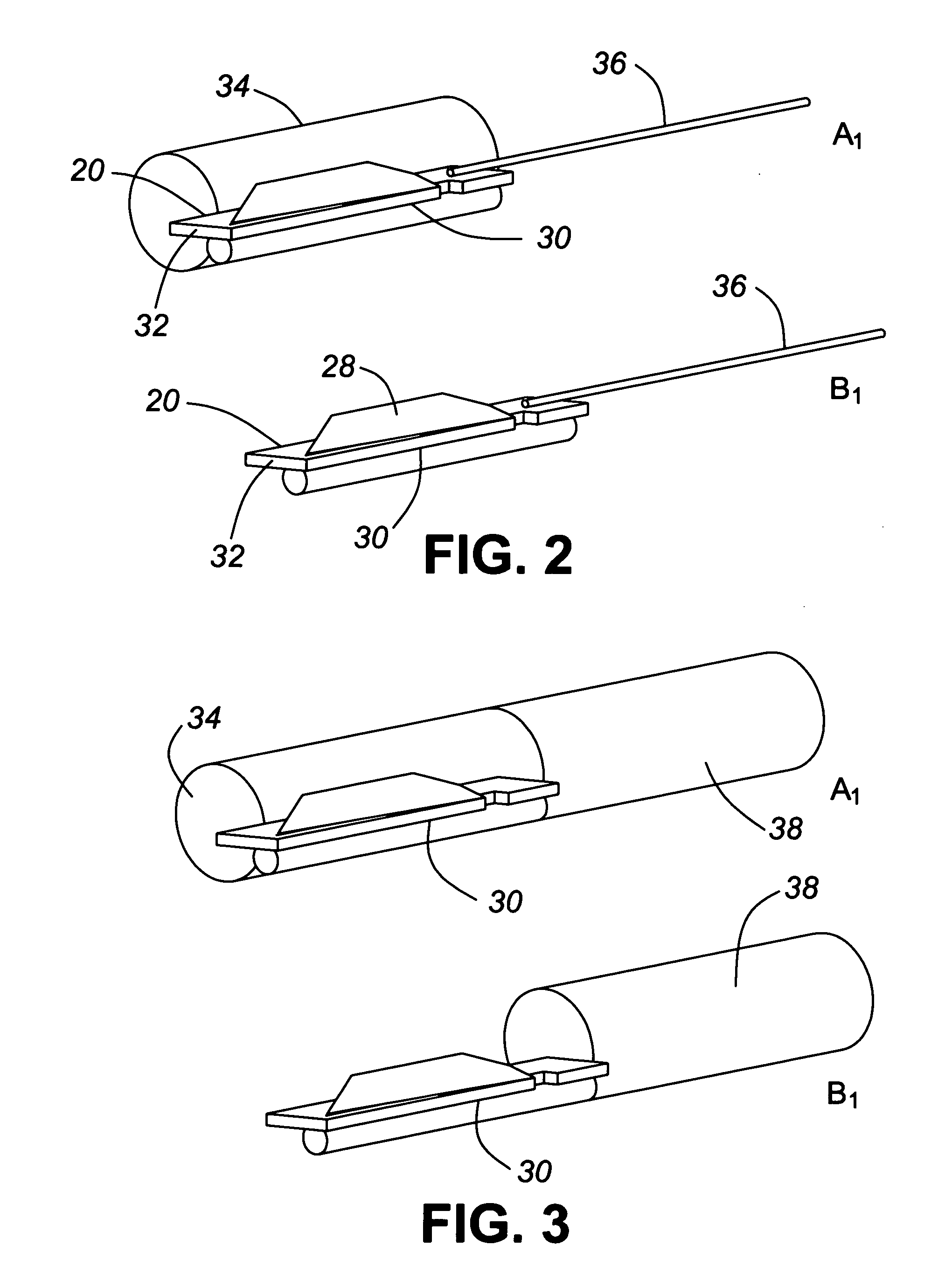
[0032]FIG. 1 illustrates a patient 10 lying on a table 12 beneath a radiation source unit 14 carried by a gantry 16 extending from a support 18 of a known kind of radiation therapy machine which irradiates a target volume on or in the patient's body in known manner. Such radiation therapy or surgery equipment is well known to those skilled in this art and so will not be described in detail herein. FIG. 1 also illustrates dosimetry apparatus for monitoring the radiation, which comprises a dosimeter 20 shown, for ease of depiction, mounted upon the patient's abdomen, and having at least one radiation detector for detecting radiation levels over a prescribed time period and providing corresponding readings or doses by way of an electronic interface unit 22, sometimes called a reader, which converts the radiation level readings into a digital format, and sends them to a computer 24 for analysis and display of the radiation dose on a display device 26, e.g., a computer monitor. The compu...


PUM


Abstract

In order to overcome or at least mitigate difficulties in compensating for movement of the target, for example a tumour during radiotherapy, a dosimetry apparatus and method embodying the present invention employ a dosimeter having at least one radiation detector and at least one magnetic position sensor located a predetermined distance apart. Radiation level readings from the radiation detector and position readings from the position sensor are monitored and correlated, conveniently according to time, to obtain the position of the radiation detector when a particular radiation level was detected.

Description

CROSS REFERENCE TO RELATED APPLICATION[0001]This application claims priority from U.S. Provisional patent application No. 60 / 904,169 filed Mar. 1, 2007; the entire contents of this prior application are incorporated herein by reference.TECHNICAL FIELD[0002]This invention relates to radiation dosimetry apparatus and methods for measuring radiation, and to dosimeters for use therein. The invention is especially, but not exclusively, applicable to radiation dosimetry apparatus and methods and dosimeters for monitoring levels of ionizing radiation during medical procedures, such as radiation therapy as used for the treatment of, for example, tumours.BACKGROUND OF THE INVENTION[0003]When ionizing radiation is being used, it is usually important to confine the radiation to the target volume and avoid irradiating surrounding material. This is especially important when using radiation therapy to treat tumours. So-called conformal radiotherapy seeks to deliver high radiation doses to the tum...

Claims


Application Information

Patent Timeline
Patent Type & Authority Applications(United States)
IPC IPC(8): H05G1/42
CPCA61N5/1001A61N2005/1072A61N2005/1055A61N5/1048A61B2034/2072A61B2090/3954
Inventor SAOUDI, ABDELHAMIDCYGLER, JOANNA EWAASHTON, ROBERT WILLIAM
Owner BEST THERATRONICS
Why Eureka
Try Eureka