Patient table comprising a positioning system and method of using such a patient table

a positioning system and patient technology, applied in the field of patient table comprising a positioning system, can solve the problems of not satisfyingly overlapping the actual acquired 2d x-ray image with the previously reconstructed 3d image, the world coordinate system and the table plate of the patient table on which the patient may be stored, and the difficulty of establishing the relationship between the world coordinate system and the table plate of the patient table,

Inactive Publication Date: 2014-07-01
KONINK PHILIPS ELECTRONICS NV
View PDF11 Cites 9 Cited by
  • Summary
  • Abstract
  • Description
  • Claims
  • Application Information

AI Technical Summary

Benefits of technology

[0011]Accordingly, there may be a need for an X-ray image acquisition arrangement and a method of using such arrangement in which a positioning of a region of interest with respect to an X-ray image acquisition device may be reliably improved.
[0029]The methods of the second or third aspect may be implemented in a patient table or an X-ray image acquisition arrangement in order to improve re-positioning capabilities or three-dimensional roadmapping capabilities, respectively.
[0035]It has been found that each of the first and second sensor arrangements may have its own advantages and deficiencies and that a positioning system comprising such two different sensor arrangements may allow improved positioning characteristics such as an improved positioning accuracy and / or an improved positioning robustness.
[0040]The proposed positioning method may be implemented into the patient table according to the first aspect of the invention by suitably adapting a control device comprised in the patient table. Thereby, an automatic positioning control may be implemented with a high positioning accuracy and a high robustness. For example, while the moving of the table plate by controlling the actuator based on the first position data may be fast, robust, easy to implement, independent of any defects of the second sensor arrangement, etc., the subsequent moving of the table plate by controlling the actuator based on a difference between the original second position data and the actual second position data may allow for a very high positioning accuracy, preferably independent of any mechanical influence.
[0041]The method for three-dimensional roadmapping according to the third aspect of the invention may benefit from the characteristics of the patient table of the first aspect of the invention in that a three-dimensional X-ray image may be acquired at a first point in time wherein the position of the table plate may be precisely recorded acquiring original position data using both, the first and second sensor arrangements, and that then, at a later second point in time and after having moved the table plate to a different position, the table plate may be moved towards the original position by controlling the actuator based on the original first position data and then acquiring one or more current two-dimensional X-ray image(s) of the object positioned on the table plate. Such current two-dimensional X-ray image(s) may be overlaid / superimposed with the originally acquired three-dimensional X-ray image in order to provide three-dimensional roadmapping for a physician. However, as the actual position of the table plate may still differ from the original position of the table plate, actual second position data of the at least one marker are acquired using the second sensor arrangement. These actual second position data may be acquired with high accuracy and may be independent of any mechanical influence onto the table plate. Based on a difference between the actual second position data and the original second position data, the three-dimensional X-ray image may be re-positioned and / or re-oriented such that an improved overlay of the transformed three-dimensional X-ray image and the currently acquired two-dimensional X-ray image(s) may be achieved.
[0042]The method according to the third aspect of the invention may be implemented in an X-ray image acquisition arrangement comprising an X-ray image acquisition device such as a C-arm system including an X-ray source and an X-ray detector. Additionally, the X-ray image acquisition arrangement comprises a patient table as defined with respect to the first aspect of the invention and a control device being adapted to control the patient table for performing the method as defined with respect to the third aspect of the invention. Using such X-ray image acquisition arrangement allows an improved 3D roadmap being essentially independent of any mechanical influence onto the table plate.

Problems solved by technology

However, a relation between the world coordinate system and the table plate of a patient table on which a patient to be examined may be stored may be more difficult to establish, due to for example an irreproducible behaviour of a patient table's positioning system.
Accordingly, it may be difficult to compare X-ray images acquired during the previous and the later image acquisition.
Or, in the example of 3D roadmapping, actually acquired 2D X-ray images may not satisfyingly overlap with a previously reconstructed 3D image.

Method used


Image

  • Patient table comprising a positioning system and method of using such a patient table
  • Patient table comprising a positioning system and method of using such a patient table
  • Patient table comprising a positioning system and method of using such a patient table

Examples


Embodiment Construction

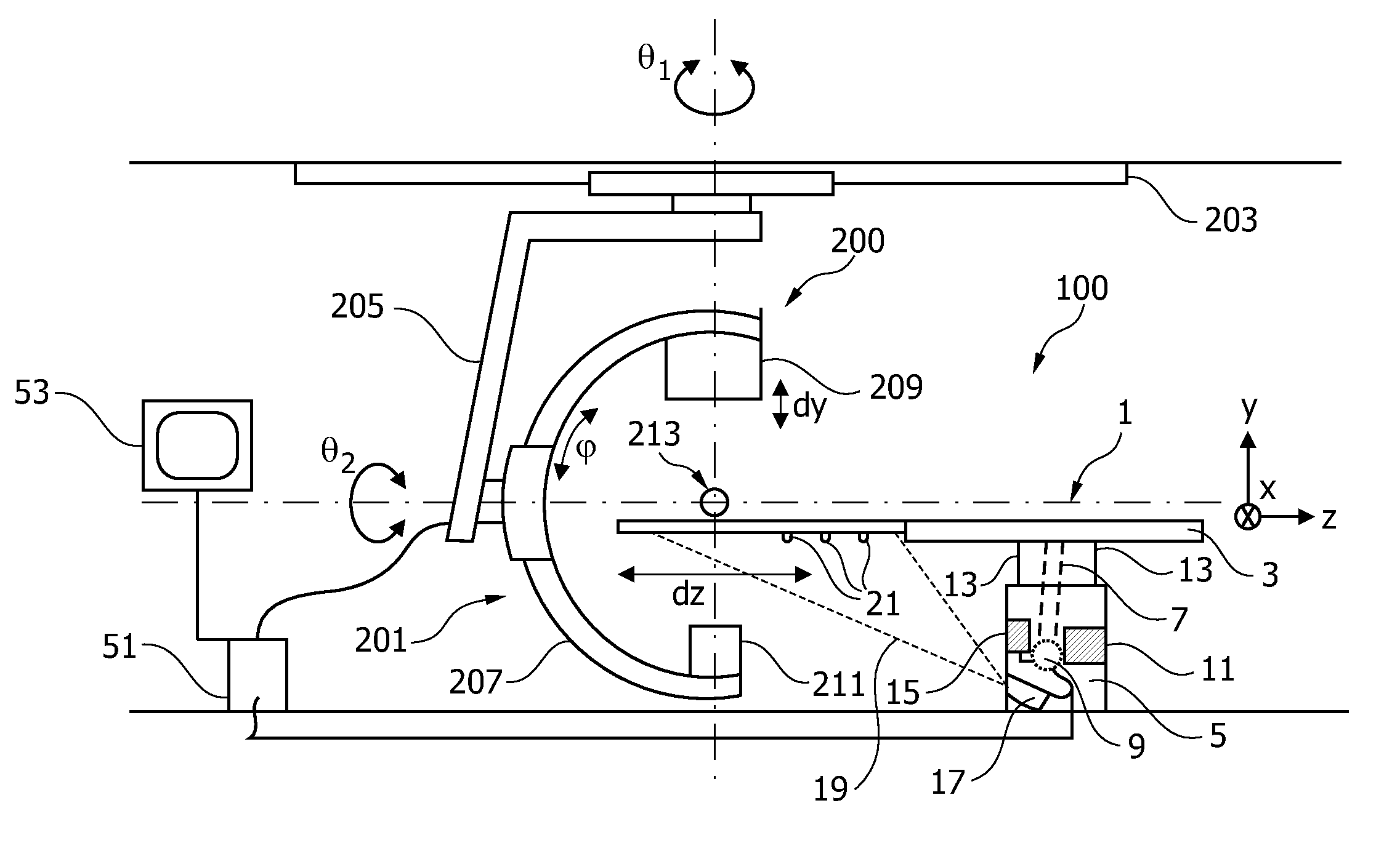
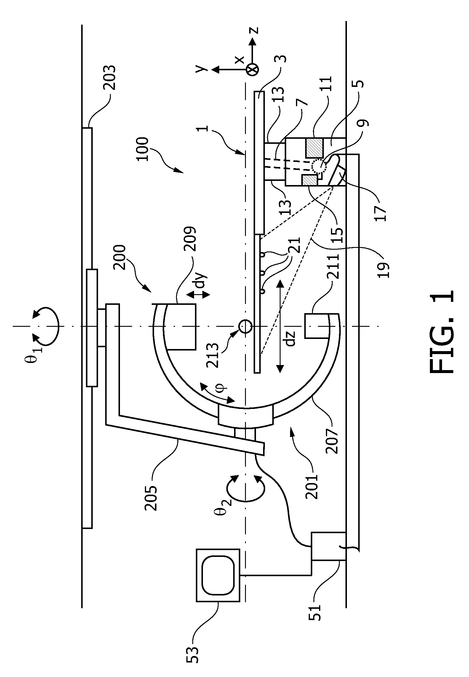
[0050]FIG. 1 shows an X-ray image acquisition arrangement 100 comprising an X-ray image acquisition device 200 and a patient table 1. The X-ray image acquisition device 200 comprises a C-arm system 201 being attached to a ceiling 203 via a movable attachment mechanism 205. The C-arm system 201 comprises a C-arm 207. On one end of the C-arm 207, an X-ray source 209 is attached. At an opposite end of the C-arm 207, an X-ray detector 211 is arranged. The C-arm system 201 may be moved translationally and rotationally along different directions or about different axes as indicated in FIG. 1 by respective arrows in order to move the C-arm along various degrees of freedom, i.e. L-arm, propeller, roll, source-imaging-distance (SID), etc. Such capability for various motions of the C-arm 207 allows to position the X-ray source 209 and the X-ray detector 211 such that X-ray images of a region of interest 213 within a patient lying on the patient table 1 can be acquired (for clarity reasons, th...


PUM


Abstract

A patient table usable in an X-ray image acquisition arrangement including a table plate connected to a table base via at least one movable joint. The table plate is movable using at least one actuator. To set or re-establish an accurate position / orientation of the movable table plate, the patient table includes a positioning system including a first sensor arrangement for providing position data of the at least one movable joint and a second sensor arrangement for providing position data of the table plate. The accurate position data provided by the second sensor arrangement may be used in a method for improved three-dimensional roadmapping.

Description

FIELD OF THE INVENTION[0001]The invention relates to a patient table comprising a positioning system. Furthermore, the invention relates to methods of using such patient table, to an X-ray image acquisition arrangement including such patient table, to a computer program element arranged to control the method of using such patient table and to a computer-readable medium having stored thereon such computer program element.BACKGROUND OF THE INVENTION[0002]X-ray image acquisition devices have been developed for various clinical applications. By emitting X-rays from an X-ray source towards an object to be examined such as for example a part of a patient and detecting the partially transmitted X-rays on the opposite side of the object, an image of the object showing its interior structure may be acquired due to the differing X-ray absorption properties for example of different types of patient tissue such as bones, muscles, etc.[0003]For example, C-arm systems have been developed which co...

Claims


Application Information

Patent Timeline
Patent Type & Authority Patents(United States)
IPC IPC(8): A61B6/04
CPCA61B6/547A61B6/12A61B6/4441A61B6/466A61B6/504A61B5/06A61B6/463A61B6/08A61B6/5235A61B6/0457A61B6/4464A61B6/0487
Inventor RUIJTERS, DANIEL, SIMON, ANNA
Owner KONINK PHILIPS ELECTRONICS NV
Why Eureka
Try Eureka