Method for determining a cardiac function

a physiological characteristic and cardiac function technology, applied in the field of physiological characteristics associated with cardiac function, can solve the problems of general difficulty in capturing and maintaining accurate pulse wave signals via the noted methods, and methods are susceptible to significant errors, so as to improve accuracy and accuracy of pulse wave velocity determination, increase in pressure and expelling of blood, and reduce pressure

Active Publication Date: 2014-09-16
CARDIAC PROFILES
View PDF10 Cites 2 Cited by
  • Summary
  • Abstract
  • Description
  • Claims
  • Application Information

AI Technical Summary

Benefits of technology

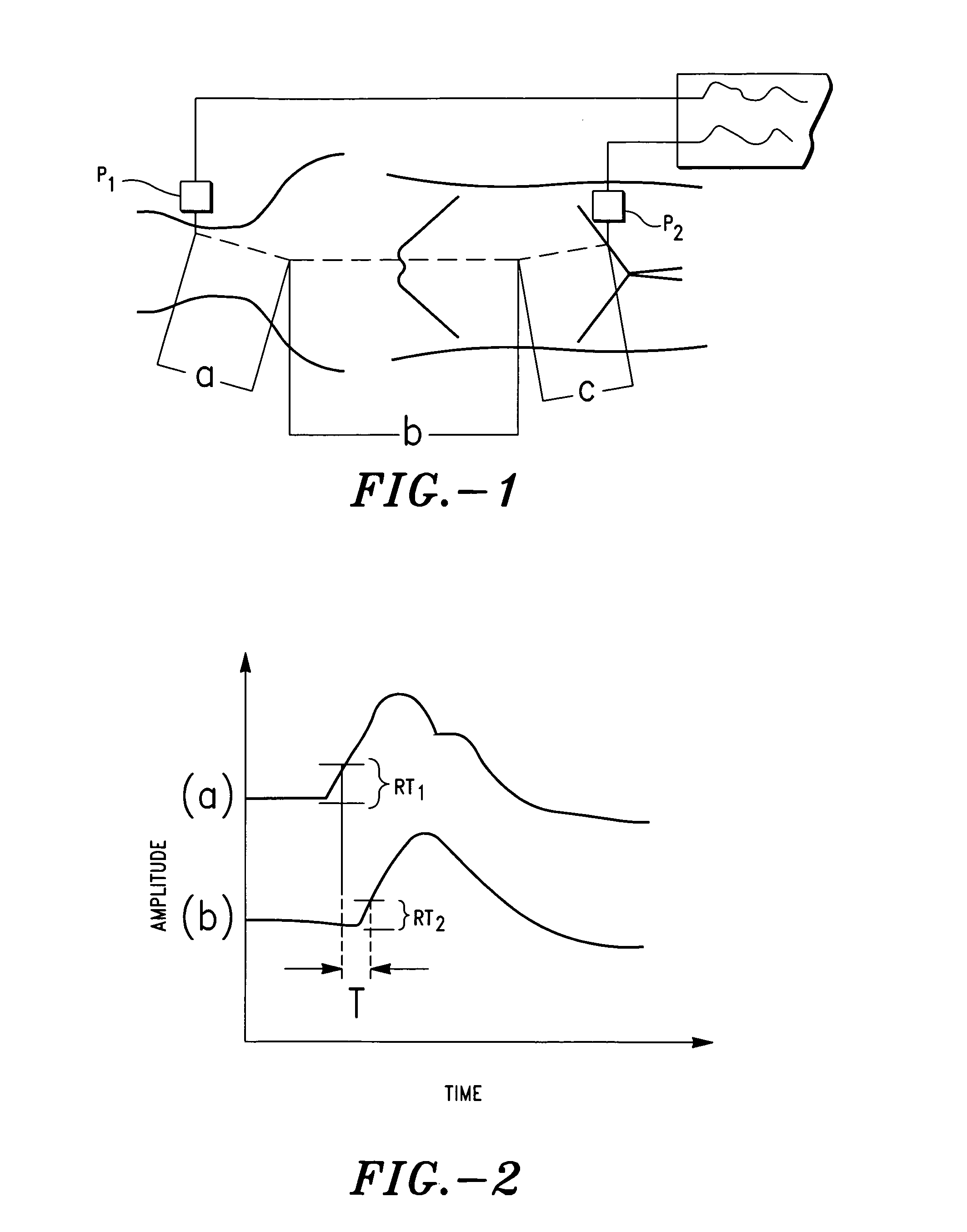
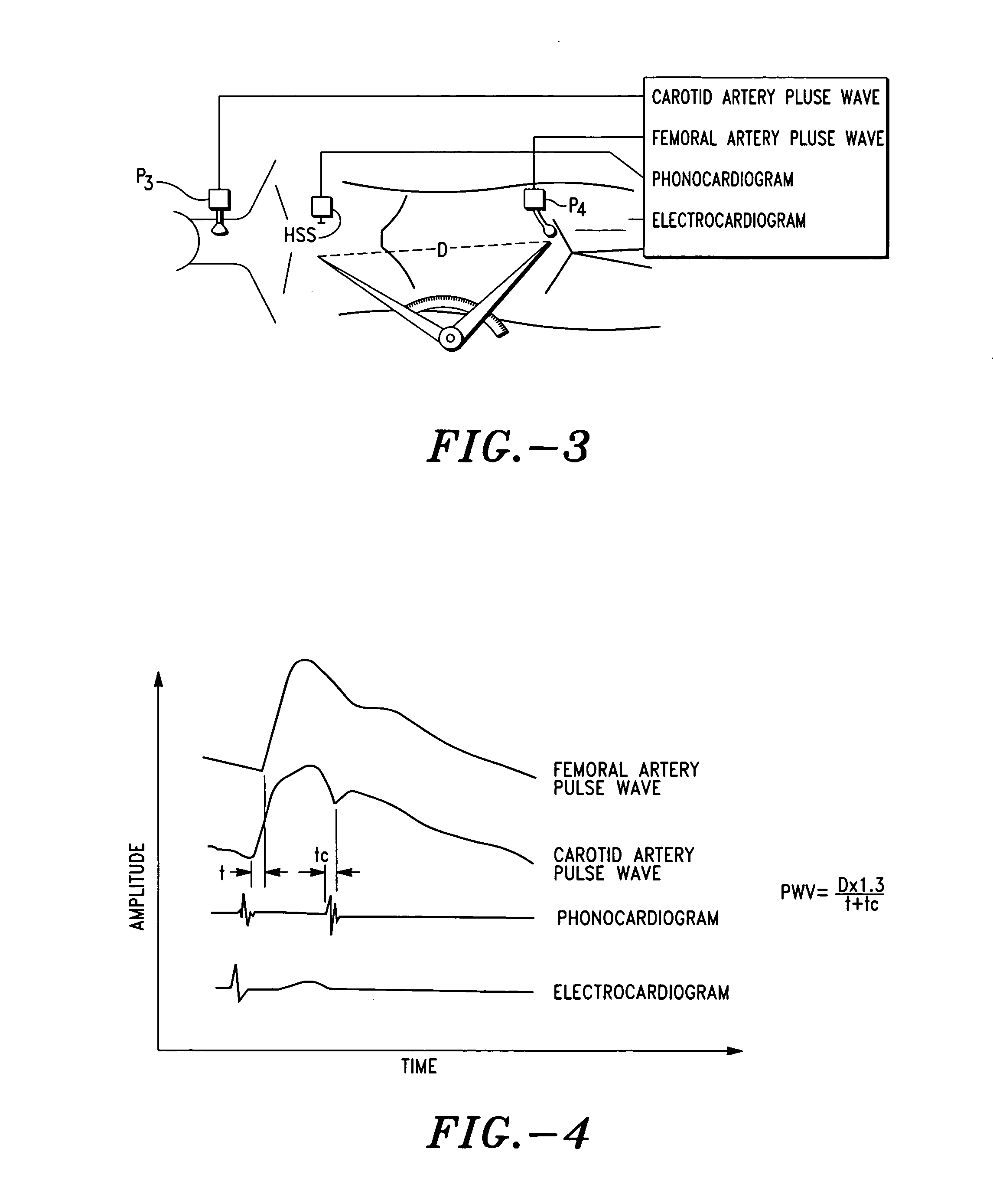
[0051]The present invention provides improved methods for determining the pre-ejection period and, hence, aortic pulse wave velocity that substantially reduces or eliminates the disadvantages and drawbacks associated with conventional methods and associated algorithms for determining the pre-ejection period and pulse wave velocity. As discussed in detail below, a key feature and, hence, advantage of the invention is that the methods and associated algorithms disclosed herein provide an accurate measure of pulse wave velocity by effectively accounting for the pre-ejection period.
[0052]As will be readily appreciated by one having ordinary skill in the art, accurate determination of pulse wave velocity is particularly beneficial in assessments of the cardiovascular system. Indeed, as discussed above, pulse wave velocity is often employed as an index of arterial elasticity and stiffness and, hence, in many instances, a predictor of coronary heart disease. Pulse wave velocity is also often employed to determine cardiac output and, hence, diagnostic information on cardiovascular performance based thereon.
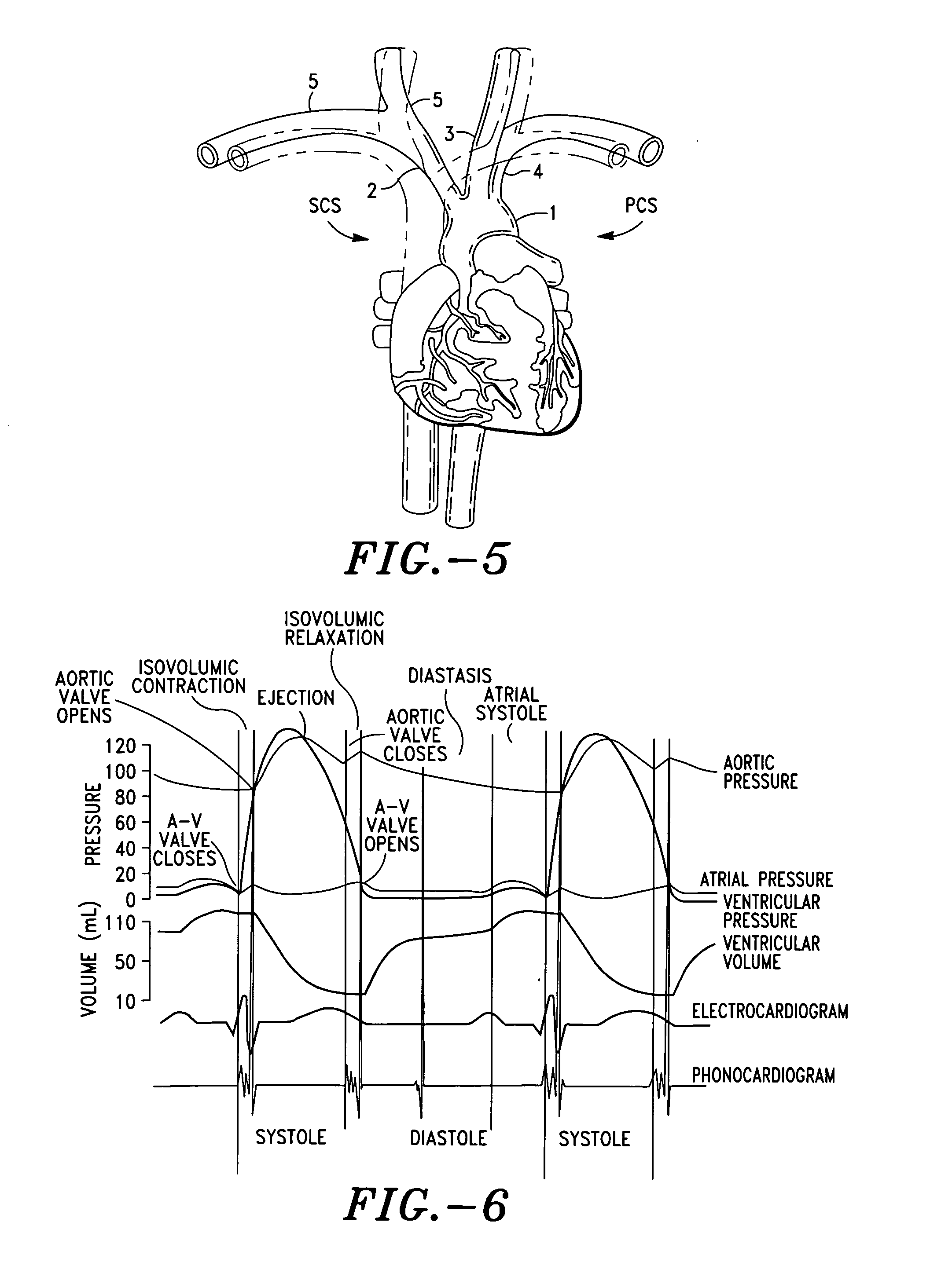
[0053]Referring now to FIG. 5, there is shown an illustration of a human heart. As illustrated in FIG. 5, functionally, the heart is divided into two sides, i.e. right and left, or sections, i.e. pulmonary and systemic circulation sections. The right or pulmonary circulation section (designated “PCS”) receives blood from the veins of the body and pumps it through the lungs. The left or systemic circulation section (designated “SCS”) receives the blood from the lungs and pumps it to the body. The blood is then collected in the veins to be returned to the right side of the heart.
[0054]The arterial system begins at the aorta 1, to which the left ventricle of the heart pumps. The aorta 1 passes down (caudad) through the body, providing arterial branches to organs, and terminates as a bifurcation, i.e. creating the iliac arteries.
[0055]The first three branches of the aorta 1 are the brachiocephalic or innominate artery 2, the left (common) carotid artery 3, and the left subclavian artery 4. The brachiocephalic artery 2 branches into the right subclavian 5 and right (common) 6 carotid arteries. These arteries provide the blood supply for the head and upper extremities.
[0056]The brachiocephalic or innominate artery 2 is the first branch of the aorta 1. The innominate artery 2, in turn, branches into the right subclavian 5 and right carotid arteries 6. In contrast, the left subclavian 4 and left carotid arteries 3 originate directly off the aortic arch. Thus, the subclavian 4 and carotid arteries 3, as well as their branches, have different paths from their counterparts on the opposite side of the body.

Problems solved by technology

There are several drawbacks and disadvantages associated with the Frank and PWV original methods.
A major drawback is that it is generally more difficult to capture and maintain an accurate pulse wave signal via the noted methods.
Although the methods disclosed in the noted references provide an effective means of determining pulse wave velocity, the methods are susceptible to significant error by virtue of the fact that the determinations of pulse transit time, which is a primary variable in pulse wave velocity equations and algorithms, fail to adequately account for the pre-ejection period (“PEP”).

Method used


Image

  • Method for determining a cardiac function
  • Method for determining a cardiac function
  • Method for determining a cardiac function

Examples


example 1

[0147]An elderly female patient is presented with the following:[0148](i) age: 92 years;[0149](ii) height: 62 inches;[0150](iii) weight: 79 Kg: and[0151](iv) arterial hypertension (systolic pressure=151 mmHg and diastolic pressure=89 mmHg).

[0152]The following basic distances were employed:

D01=D0x+Dx1=8.00 cm+13.00 cm=21 cm;  (i)

D02=D0x+Dx2=8.00 cm+74.00 cm=82 cm; and  (ii)

D12=D02−D01=82.00 cm−21.00 cm=61 cm.  (iii)

[0153]The following measurements were also determined:[0154](i) D0x=8.00 cm;[0155](ii) Dx1=13.00 cm; and[0156](iii) Dx2=74.00 cm.

[0157]From measured patient data the following was also provided:[0158](i) PDDigit=0.1984 seconds; and[0159](ii) PDEar=0.1627 seconds.

[0160]Using a value of 0.88 for ratio parameter αv, pulse wave velocity (PWV) and pre-ejection period (PEP) is then determined, as set forth above, i.e.

[0161]PWVCentral=αv×PWVPeripheral=D12×αv(PDDigit-PDEar)=61⁢⁢cm×0.88(0.1984sec-0.1627sec)=1627⁢⁢cmsecP⁢⁢E⁢⁢P=PDEar-(PDDigit-PDEar)*D01(D02-D01)*αage=0.1627sec-(0.198...

example 2

[0162]A young normotensive female patient is presented with the following:[0163](i) age: 42 years;[0164](ii) height: 63 inches;[0165](iii) weight: 79 Kg: and[0166](v) arterial hypertension (systolic pressure=130 mmHg and diastolic pressure=7 mmHg).

[0167]The following basic distances were employed:

D01=D0x+Dx1=6.35 cm+13.97 cm=20.32 cm;  (i)

D02=D0x+Dx2=6.35 cm+80.01 cm=86.36 cm; and  (ii)

D12=D02−D01=86.36 cm−20.32 cm=66.04 cm.  (iii)

[0168]The following measurements were also determined:[0169](i) D1=6.35 cm;[0170](ii) D2=13.97 cm; and[0171](iii) D4=80.01 cm.

[0172]From measured patient data the following was also provided:[0173](i) PulseDelayDigit=0.1942 seconds; and[0174](ii) PulseDelayFacial=0.1472 seconds.

[0175]Using a value of 0.88 for ratio parameter αv, pulse wave velocity (PWV) and pre-ejection period (PEP) is then determined, as set forth above, i.e.

[0176]PWVCentral=αv×PWVPeripheral=D12×αv(PDDigit-PDEar)=66.04⁢⁢cm×0.88(0.1942sec-0.1472sec)=955⁢⁢cmsecP⁢⁢E⁢⁢P=PDEar-(PDDigit-PDEar)...


PUM


Abstract

A method for determining a cardiac function, comprising (i) determining base anatomical characteristics associated with the subject, (ii) determining pulse delay to a first body site (PD01) and a second body site (PD02) as a function of the anatomical characteristics, wherein the distance via the arterial tree from the aortic valve to the first body site (PD01) is different than the arterial tree distance from the aortic valve to the second body site (PD02), (iii) determining pulse wave velocity between the first body site and the second body site (PWV12), (iv) determining pulse wave velocity between the aortic valve and the first body site (PWV01) as a function of PWV12, and the anatomical characteristics; and (v) determining the pre-ejection period (PEP) as a function of PD01 and PWV01.

Description

FIELD OF THE PRESENT INVENTION[0001]The present invention relates generally to methods of determining physiological characteristics associated with cardiac function. More specifically, the invention relates to improved methods for determining the pre-ejection period and pulse wave velocity.BACKGROUND OF THE INVENTION[0002]The study of the performance and properties of the physiology (including notably the cardiovascular system) of a living subject has proven useful for diagnosing and assessing any number of conditions or diseases within the subject. The performance of the cardiovascular system, particularly the heart, has characteristically been measured in terms of several pertinent parameters, such as pulse wave velocity, pulse transit time, stroke volume and cardiac output.[0003]A key cardiovascular parameter (or physiological characteristic) is pulse wave velocity, i.e. the speed at which a pressure wave propagates throughout the arterial tree, or aortic pulse wave velocity, i.e...

Claims


Application Information

Patent Timeline
Patent Type & Authority Patents(United States)
IPC IPC(8): A61B5/02
CPCA61B5/026A61B5/02125
Inventor VOSS, GREGORY I.PERRY, JAMES M.CLINTON, III, RANKIN A.STERLING, BERNHARD B.LAWRENCE, ANDREW R
Owner CARDIAC PROFILES
Why Eureka
Try Eureka