Cardiac image processing and analysis

a cardiac image and processing technology, applied in the field of visualizing a cardiac image, can solve the problems that visualizations do not provide an efficient way to inspect the myocardium in detail, and achieve the effects of improving motion compensation, and reducing the number of visualization errors

Active Publication Date: 2014-09-30
KONINK PHILIPS ELECTRONICS NV
View PDF18 Cites 2 Cited by
  • Summary
  • Abstract
  • Description
  • Claims
  • Application Information

AI Technical Summary

Benefits of technology

[0020]The motion-compensating means may be arranged for employing a rigid or affine transformation to transform a cardiac image into a motion-compensated image. Such a rigid or affine transformation is known per se in the art. By limiting the transformation to rigid or affine transformations, most ‘healthy’ kinds of cardiac motion can be compensated. Pathologic, or irregular cardiac motion may not be compensated by such transformations. Asynchronies, for example, would normally not be compensated by using a rigid or affine transformation. Consequently, these pathologic or irregular cardiac motions remain in the motion-compensated images as residual motion. In combination with the resampling and visualization modes set forth, the residual motion can be identified by a user relatively efficiently, and thus the pathologic or irregular cardiac motion or asynchronies can be identified relatively efficiently.
[0022]Indicating means may be provided for enabling a user to indicate a region comprising residual motion; and the motion-compensating means may be arranged for further motion-compensating a region indicated by the user. The visualization of a cardiac image sequence as set forth allows easy spotting of residual motion. By enabling the user to identify this residual motion to the system, the system can apply further motion compensation specific to this region.
[0023]The indicating means may be arranged for further enabling the user to indicate a direction of the residual motion, the further motion-compensating means being arranged for compensating the region indicated by the user according to a direction of the residual motion indicated by the user. By using the direction, the motion compensation is improved further.

Problems solved by technology

However, these visualizations do not provide an efficient way to inspect the myocardium in detail.

Method used


Image

  • Cardiac image processing and analysis
  • Cardiac image processing and analysis
  • Cardiac image processing and analysis

Examples


Embodiment Construction

[0040]Cardiac tissue motion fields can be extracted, for example, directly from the imaging mode (Doppler US or specialized MRI modes) or by using a rigid or non-rigid registration technique, for example as described in Ledesma-Carbayo et al. However, several difficulties have to be solved in order to correct and assess the validity of the estimated motion-field.

[0041]The acquired image contrast may be low in the region where motion has to be assessed. Examining the motion-compensated image sequence may be a tedious and lengthy task when using 3D time sequences (4D visualization).

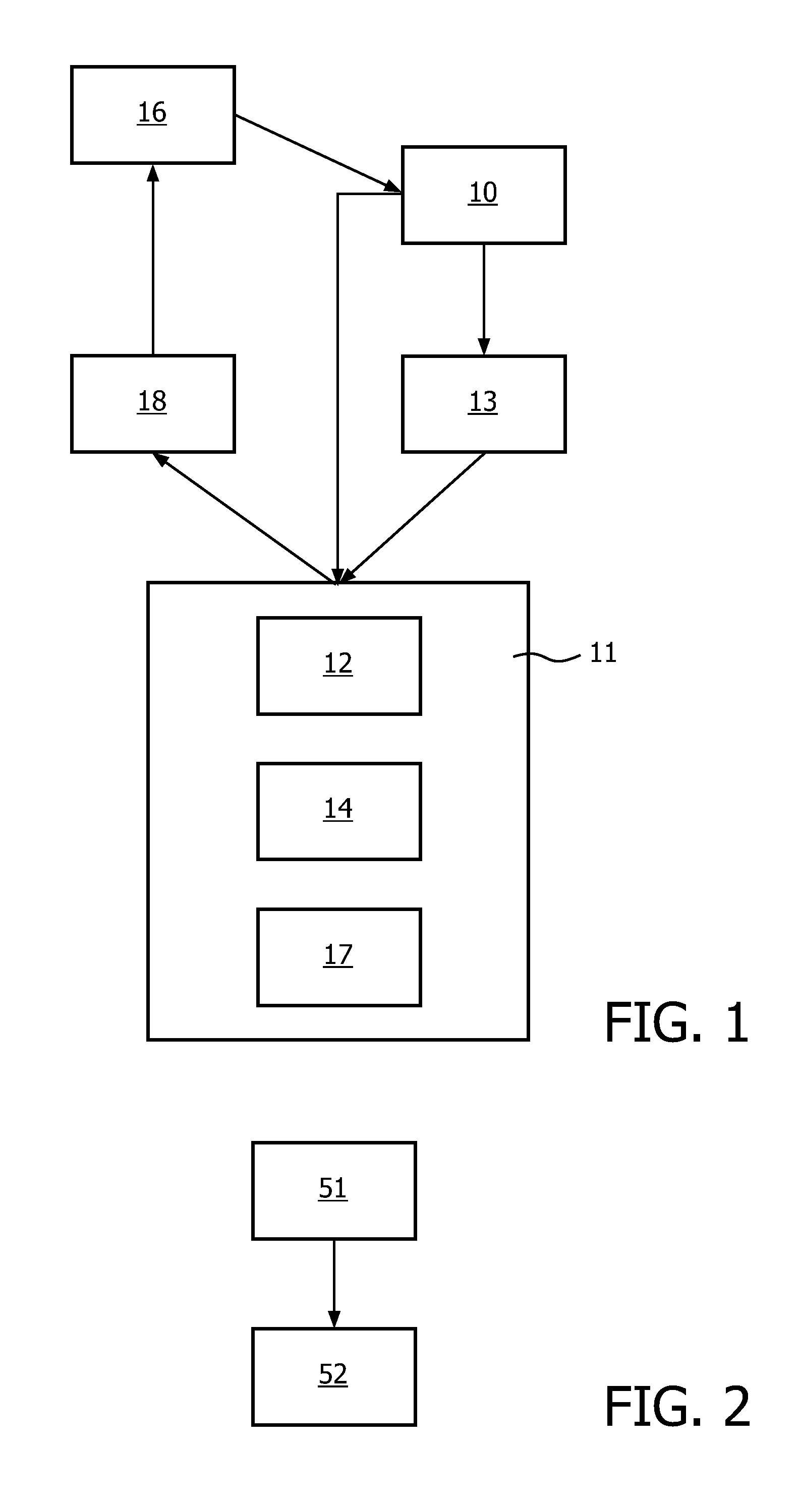
[0042]FIG. 1 illustrates a system for visualizing a myocardium represented by a cardiac image associating intensity levels with three-dimensional points in a volume. Such a cardiac image is known as a volumetric cardiac image. The system may comprise a computer system including a central processor unit, memory, and storage means, as known in the art. Some or all of the means and modules described herein can...


PUM


Abstract

A system for visualizing a myocardium represented by a cardiac image comprises a resampling means and a visualizing means. The resampling means resamples the intensity levels at sampling points on a plurality of curved surfaces, each curved surface enclosing at least part of a heart cavity and zero or more of the plurality of curved surfaces and being enclosed by the remaining curved surfaces of the plurality of curved surfaces, the plurality of curved surfaces together covering a hollow region in the cardiac image, the hollow region comprising the outer cavity walls of a group of at least one heart cavity. The visualizing means is arranged for visualizing at least part of at least one of the plurality of curved surfaces, using resampled intensity levels obtained from the resampling means. The group of at least one heart cavity may be the left atrium alone. It may also be the complete heart.

Description

FIELD OF THE INVENTION[0001]The invention relates to visualizing a cardiac image. The invention also relates to motion compensation of a sequence of cardiac images. The invention also relates to strain imaging.BACKGROUND OF THE INVENTION[0002]The motion of cardiac tissues can be assessed by viewing medical images of the cardiac tissues, using one or more different 2D and 3D imaging modes (in particular tagged or untagged cardiac MRI, and / or different modes of ultrasound imaging). From such dynamic imaging, one can extract tissue velocity, strain and / or strain-rates. Such strain and strain rates can be visualized using parametric images, wherein the intensities associated with positions represent parameter values, such as strain or strain rate. Such parametric imaging techniques can provide valuable diagnostic information relating, among others, to hypokinesy, akinesy, diskinesy of certain part of the myocard, or myocardial asynchronism. However, the validity of this parametric imagi...

Claims


Application Information

Patent Timeline
Patent Type & Authority Patents(United States)
IPC IPC(8): G06K9/00A61B6/00A61B8/08G06T7/00
CPCA61B8/08G06T7/0083A61B6/503G06T2207/30048G06T7/12
Inventor ALLAIN, PASCALMAKRAM-EBEID, SHERIFSOMPHONE, OUDOM
Owner KONINK PHILIPS ELECTRONICS NV
Why Eureka
Try Eureka