Microsurgery simulator

a micro-surgery and simulator technology, applied in the field of micro-surgery simulators, can solve the problems of not well representing the actual human anatomy, inability to support a variety, and high cos

Inactive Publication Date: 2015-07-28
SIMQUEST
View PDF9 Cites 3 Cited by
  • Summary
  • Abstract
  • Description
  • Claims
  • Application Information

AI Technical Summary

Benefits of technology

[0010]One or more embodiments of the simulator provides a means for surgeries performed under optical magnification, either by a base supported surgical microscope or head-worn lopes, to be practiced by replicating the response of tissue being acted upon by microsurgical tools held by the learner, typically one in each hand. The handles of the same tools that are used in real procedures are held and manipulated by the learner / user. These tools are connected to haptic interface devices that track the learner's positioning and orientation of the tools as well their manipulation of the tool mechanism(s) such as jaws. This information is fed into a real-time physics simulation of the interaction of the tools with appropriate computational models of the tissue at the surgical site of interest. The resulting stress / strain / deformation of the tissue is displayed to the learner and the accumulated forces and moments upon the surgical tools are sent out to the haptic devices so that the learner feels the resulting interaction they have produced in manipulating the tissue. The visual display of the tissue's response to manipulation can be presented on either a surrogate surgical microscope or a head-mounted display that approximates a pair of surgical lopes. In this way, the learner sees and feels the resulting response of the tissue to their actions with the tools. Combining this simulation with models of the specific surgical sites and means to objectively observe and assess the quality of the learner's performance of actions relative to quantitatively defined standards provides the learner with a means to experience surgical situations and receive feedback about their performance without the need for an actual operating room, patient, or skilled instructor / mentor. The simulator can optionally be combined with physical models / surrogates that approximate anatomical surfaces so that the learner can place their hands upon the model for finger- / touch-based interaction in addition to tool-based interaction. These physical models / surrogates can be observed with cameras and placed upon load sensing devices so that the learner's hand actions upon the anatomy can be measured and input into the simulation. The feel of the learner's contact with the physical anatomy is provided by the passive feel of the anatomy models which are typically built from a form of soft material, such as silicone rubber derivatives, designed to approximate the appearance and feel of human tissue. The measured hand actions upon the anatomy are fed into the simulation just like the tool-based actions. In addition, in combination with the physical anatomy option, rather than using haptic interface devices, real surgical tools being spatially tracked can be used and may decrease the cost of the hardware in the simulator. There are a number of combinations of interface technologies that can be used to create the microsurgery training system / simulator. These different interface technologies may be adapted for use with the same general computational physics simulation core software. Measurement, simulation, load feedback, and visual display may be designed to provide the learner with millimeter-scale experiences similar to what occurs in real life microsurgery procedures.
[0013]According to one or more of these embodiments, the eye piece includes a dual-eye eye piece, and the mobile device is configured to render the real time computer graphics generated video representation in 3D as left and right real time computer graphics generated video representations that are viewable through left and right sides, respectively, of the dual-eye eye piece to provide depth of view.

Problems solved by technology

This approach is costly since it slows down the pace of operations, dangerous since it exposes patients to less skilled operators, limiting since only the patients with whatever maladies they might have are available for the learner to experience, and inefficient since learning can only occur when a staffed operating room, a patient, and a skilled mentor are available.
Each has distinct disadvantages in that they: do not well represent the actual human anatomy, cannot support a variety of variations in geometry, mechanical properties, or systemic physiologic response to interaction, are often either single- or limited-time use, require specialized facilities and personnel to set up and maintain during training sessions that learners must come to, tend to be costly, and / or require experienced mentors to observe learner actions and provide feedback

Method used


Image

  • Microsurgery simulator
  • Microsurgery simulator
  • Microsurgery simulator

Examples


Embodiment Construction

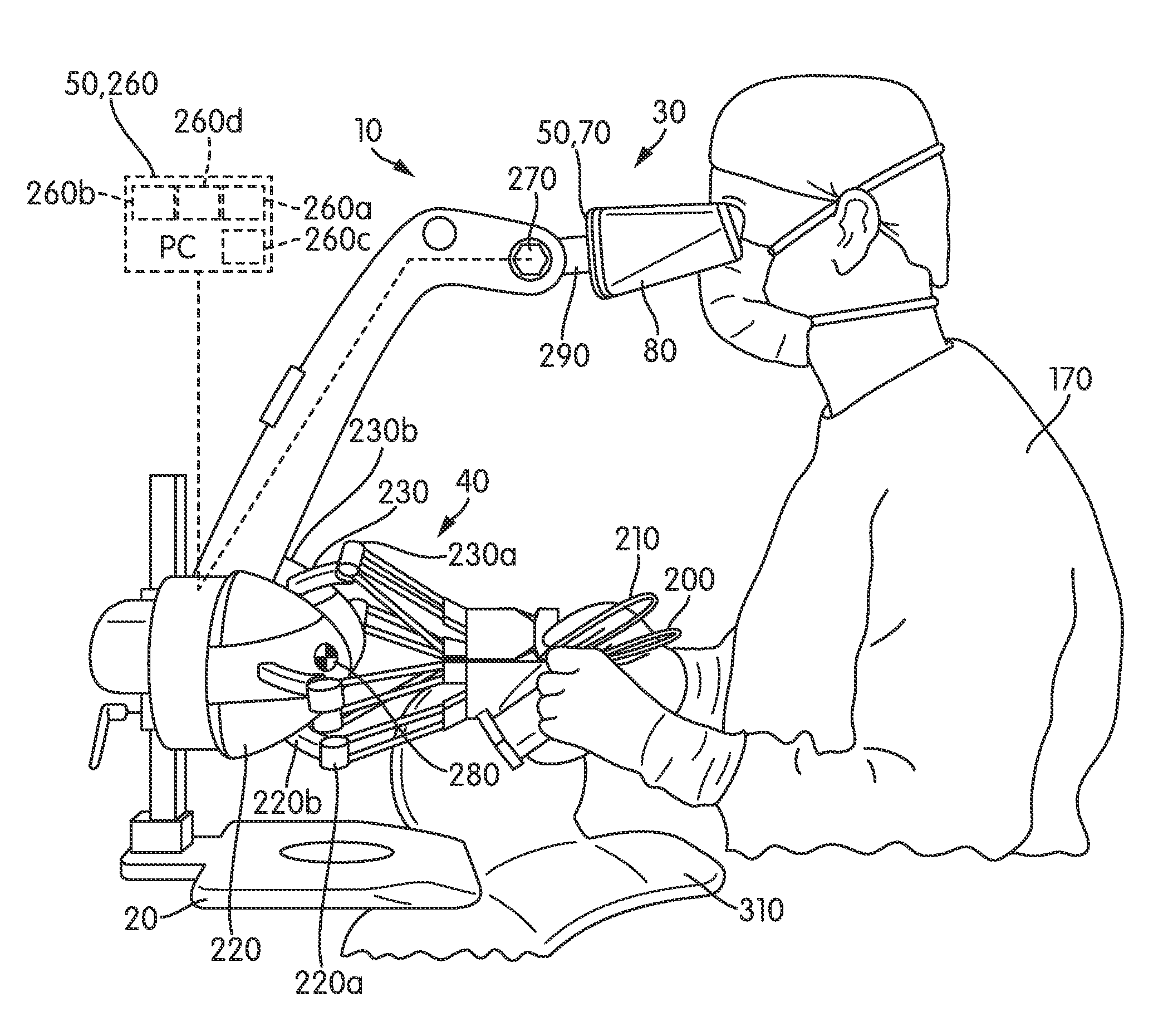
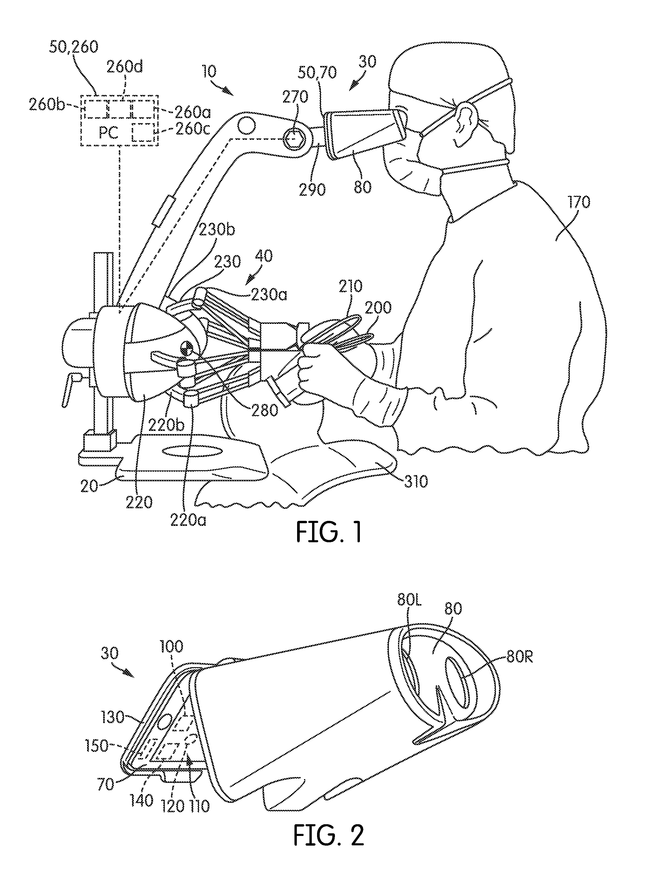
[0008]There are simulators available for minimally invasive surgical (MIS) skills training Key aspects of various conventional simulators in contrast to one or more embodiments of microsurgical simulators according to the present invention are that the real-life microsurgical procedures are performed with the surgeon's / clinician's neither having direct contact with the patient nor looking at the patient (they typically look at a monitor displaying an indirect view of the surgical site taken by a tool such as an endoscope or fluoroscope) and that the computational means used to model tool-tissue interaction are not designed to replicate real-life behavior to a provable objective metric compared to real life. Various conventional MIS simulators also do not operate on the small motion scale of microsurgical procedures. Finally, the software in various conventional MIS simulators is typically rigidly configured to work with only specific physical interfaces so that they cannot be reconf...


PUM


Abstract

A microsurgery simulator simulates various microsurgical procedures (e.g., nerve reapproximation, ear drain deployment) that utilize a surgical microscope. A surrogate surgical microscope includes a mobile device and an eye piece. A physical surrogate surgical interface represents an interface between a user and a simulated surgical scenario. Sensors sense the user's manipulation of the surrogate surgical interface. A surgical simulation generator generates a real time 3D surgical simulation based on the sensed manipulation. The generator renders the real time surgical simulation into a real time computer graphics generated video representation that is displayed on a screen of the mobile device. A processor of the mobile device is configured to perform at least a portion of a computational analysis that is used to generate the real time computer graphics generated video representation.

Description

CROSS REFERENCE[0001]This application claims the benefit of priority from U.S. Provisional Application Ser. No. 61 / 605,405, filed Mar. 1, 2012, titled “Microsurgery Simulator,” the entire contents of which are hereby incorporated by reference herein.STATEMENT REGARDING FEDERALLY SPONSORED RESEARCH AND DEVELOPMENT[0002]The U.S. Government has a paid-up license in this invention and the right in limited circumstances to require the patent owner to license others on reasonable terms as provided for by the terms of NIH Contract No. NIH NIDCR SBIR R43DE021641-01.BACKGROUND OF THE INVENTION[0003]1. Field of the Invention[0004]The present invention relates generally to surgical simulators.[0005]2. Description of Related Art[0006]Learning microsurgical skills is a hands-on process that typically requires numerous hours of practice in the operating room with real patients. This approach is costly since it slows down the pace of operations, dangerous since it exposes patients to less skilled ...

Claims


Application Information

Patent Timeline
Patent Type & Authority Patents(United States)
IPC IPC(8): G09B7/04G09B23/28
CPCG09B23/28
Inventor MEGLAN, DWIGHT A.KING, DANIEL R.DVORNIK, ALBERTLENOIR, JULIEN
Owner SIMQUEST
Why Eureka
Try Eureka