

Method for preparing cell standard
a cell standard and histological examination technology, applied in the field of histological examination methods, can solve the problems of difficult to establish a consistent quality of staining samples, difficulty in accurately determining the thickness of sectioned tissue samples, and inability to accurately evaluate the intensity of staining based on visual estimation
- Summary
- Abstract
- Description
- Claims
- Application Information
AI Technical Summary
Benefits of technology
Problems solved by technology
Method used
Image

Examples
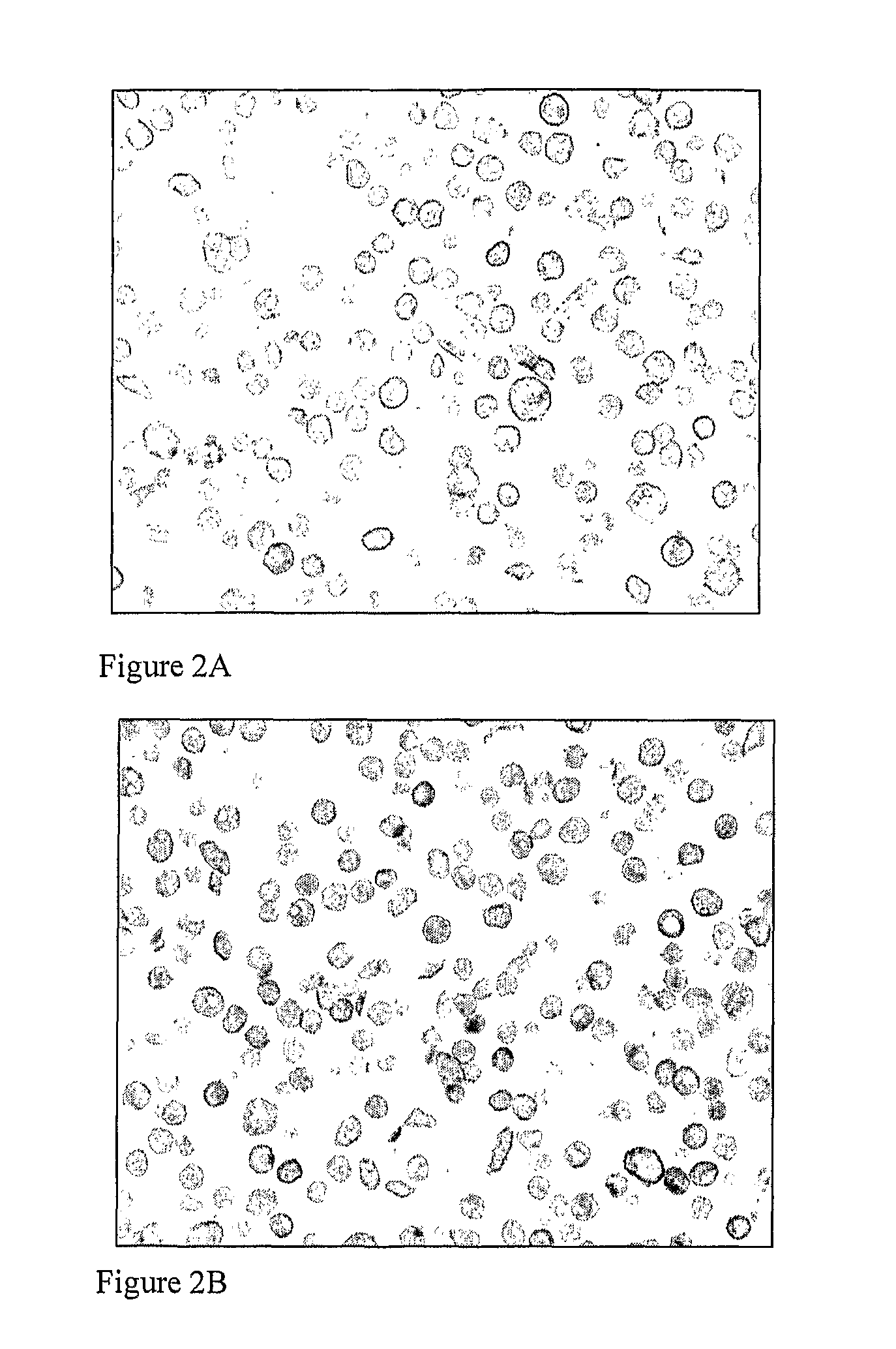
example 1
Preparation of Her 2 Control Slides
The control slides are prepared using the following cell lines:
[0067]Negative control cell line MDA-MB-231 (obtained from ECACC—ECACC No. 92020424), positive 1+ control cell line MDA-MB-175-VII (obtained from ATCC—ATCC Catalogue No. HTB-25), positive 2+ control cell line MDA-MB-453 (obtained from ATCC—ATCC Catalogue No. HTB-131) and positive 3+ control cell line SK-BR-3 (obtained from ATCC—ATCC Catalogue No. HTB-30).
[0068]Cell lines were obtained from the European Collection of Cell Cultures (ECACC) and the American Type Culture Collection (ATCC)—as detailed in the paragraph above—and grown using standard media and techniques to produce approximately 1-2×10⁸ cells. The cell growth is divided into three batches of approximately 3.3-6.6×10⁷ cells. Cultured cells scraped from the surface of the appropriate number of flasks are spun down and resuspended in formalin and fixed. Each block is paraffin embedded, cut into sections and mounted on a charged g...
example 2
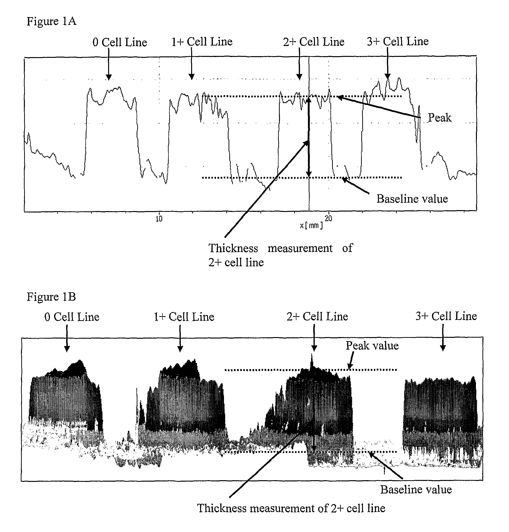
Materials and Methods
[0079]Analysis of cell line section thickness was performed in three stages. For each stage, sections of control cells lines were cut from formalin fixed paraffin embedded (FFPE) blocks using a calibrated automated microtome (RM2255, Leica Microsystems). The blocks contained four FFPE cores: MDA-MB-231 (0) (European Collection of Cell Cultures—Catalogue No. 92020424), MDA-MB-175-VII (1+) (American Type Culture Collection—ATCC Catalogue No. HTB-25), MDA-MB-453 (2+) (ATCC, Catalogue No. HTB-131) and SK-BR-3 (3+) (ATCC, Catalogue No. HTB-30). The cell lines were grown using standard cell culture techniques, fixed in formalin, re-suspended in an agarose matrix and then processed on an automated Peloris (Leica Microsystems) tissue processor by dehydration in graded alcohols (70%, 90%, 100%) and cleared in xylene, before impregnation and embedding in paraffin wax.
[0080]The breast tumour tissue microarrays slides were purchased from Stretton Scientific, UK.
Stage 1. Tes...
PUM
| Property | Measurement | Unit |
|---|---|---|
| thickness | aaaaa | aaaaa |
| thickness | aaaaa | aaaaa |
| thickness | aaaaa | aaaaa |
Abstract
Description
Claims
Application Information