
Devices, systems and methods for using and monitoring medical devices

Active Publication Date: 2016-10-27
CANARAY MEDICAL INC
View PDF6 Cites 87 Cited by
  • Summary
  • Abstract
  • Description
  • Claims
  • Application Information

AI Technical Summary

Benefits of technology

The patent describes a device that can detect movement or motion of a medical device. This helps to prevent problems during surgery or medical examinations. The device uses sensors and accelerometers to accurately measure the movement and can detect if the device is moving or if there is movement between the device and the surrounding tissue. This information helps to reduce the likelihood of the device being placed incorrectly, malfunctioning, or causing side effects during daily activities after the patient returns home.

Problems solved by technology

Unfortunately, various complications may arise during insertion of the medical device or implant (whether it is an open surgical procedure or a minimally invasive procedure).
This can however be difficult to do during the procedure itself, making corrective adjustments difficult.
In addition, a patient may experience a number of complications post-procedure.
While some of these problems can be addressed with pharmaceutical products and / or further surgery, they are difficult to predict and prevent; often early identification of complications and side effects is difficult or impossible.
Sudden increases in strain may indicate that too much stress is being placed on the medical device, which may increase damage to the body and / or breakage, cracking and / or damage to the device.

Method used


Image

  • Devices, systems and methods for using and monitoring medical devices
  • Devices, systems and methods for using and monitoring medical devices
  • Devices, systems and methods for using and monitoring medical devices

Examples


embodiment 1

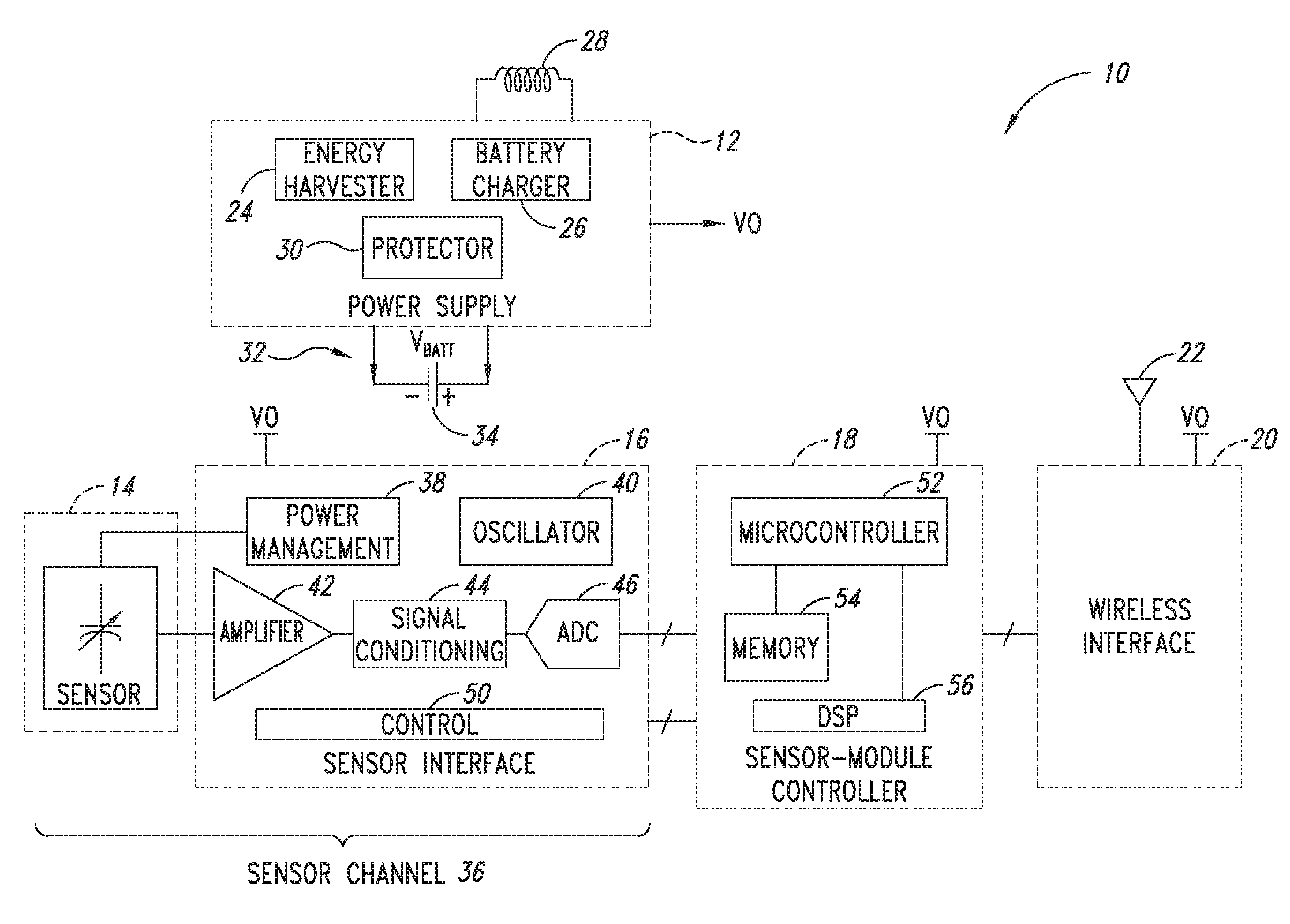
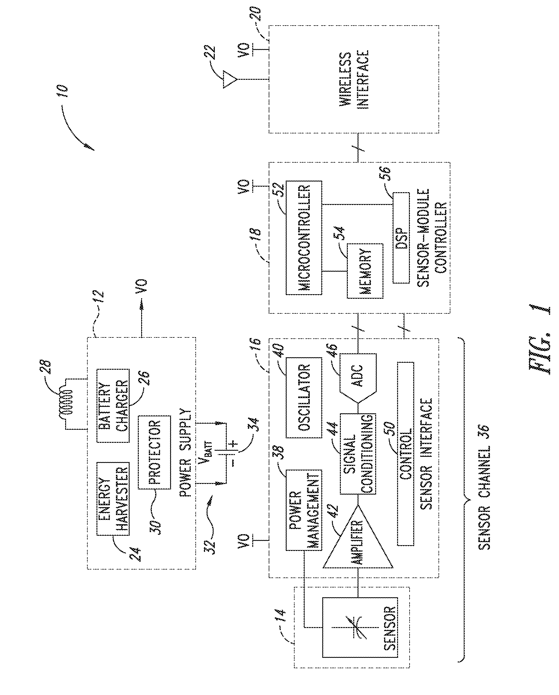
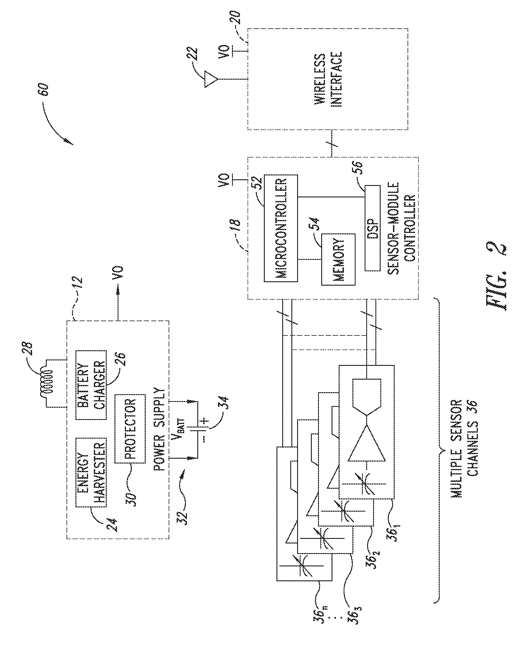
[0683]2. The sensor module of embodiment 1 wherein the sensor channel includes a sensor and a sensor channel coupled to the sensor and to the communication interface.

[0684]3. The sensor module of embodiments 1 or 2 wherein the sensor includes one or more of the following sensors: a global-positioning-system (GPS), accelerometer, Hall-effect, electrical, magnetic, thermal, pressure, radiation, optical, quantity-differential, capacitive, inductive, and time.

[0685]4. The sensor module of any one of embodiments 1 to 3 wherein the sensor is a microelectromechanical sensor. (MEMS).

[0686]5. The sensor module of any one of embodiments 1 to 4 wherein said sensor module includes one or more of the following sensors: fluid pressure sensors, fluid volume sensors, contact sensors, position sensors, pulse pressure sensors, blood volume sensors, blood flow sensors, chemistry sensors, metabolic sensors, accelerometers, mechanical stress sensors and temperature sensors. Within preferred embodiments ...

embodiment 13

[0695]14. The medical device , wherein said medical device is a cardiovascular device, orthopedic device, spinal device, intrauterine device, cochlear implant, aesthetic implant, dental implant, medical polymer or artificial eye lense.

embodiment 14

[0696]15. The medical device wherein said cardiovascular device is an implantable cardioverter defibrillator, pacemaker, stent, stent graft, bypass graft, catheter, or heart valve

[0697]16. The medical device according to embodiment 14, wherein said orthopedic device is a cast, brace, tensor bandage, support, sling, tensor bandage, hip or knee prosthesis, orthopedic plate, bone screw, spinal cage, artificial disc, orthopedic pin, intramedullary device, K-wire, or orthopedic plate. Within one embodiment the medical device is a tibial extension on a total arthroplastic joint (e.g, total hip or knee joint).

[0698]17. The medical device according to embodiment 14, wherein said medical polymer is a biodegradable polymer.

[0699]18. The medical device according to embodiment 14, wherein said medical polymer is a non-biodegradable polymer.

[0700]19. The medical device according to embodiment 14, wherein said medical polymer is a polymethylmethacrylate, a methylmethacrylate-styrene copolymer, f...


PUM


Abstract

Medical devices are provided, comprising a medical device and a sensor.

Description

CROSS-REFERENCE TO RELATED APPLICATIONS[0001]This application is a continuation-in-part of International Application No. PCT / US2015 / 050789, filed Sep. 17, 2015, which claims the benefit under 35 U.S.C. §119(e) of U.S. Provisional Patent Application No. 62 / 051,855 filed Sep. 17, 2014, and U.S. Provisional Patent Application No. 62 / 184,820 filed Jun. 25, 2015, all applications are incorporated herein by reference in their entireties.FIELD OF THE INVENTIONTechnical Field[0002]The present invention relates generally to medical devices, and more specifically, to devices and methods for monitoring the placement, efficacy, durability and performance of a wide variety of temporary and / or permanently implantable medical devices.BACKGROUNDDescription of the Related Art[0003]Medical devices and implants have become common-place in modern medicine. Typically, medical devices and implants are manufactured to replace, support, or enhance an anatomical or biological structure. Examples of medical ...

Claims


Application Information

Patent Timeline
IPC IPC(8): A61B5/00A61B5/021A61B5/029A61B5/026A61B5/03A61N1/36A61N1/37A61N1/39A61F2/16A61F2/00A61F2/07A61F2/32A61F2/38A61F5/01A61F5/055A61B17/74A61F2/24A61C8/00A61B5/01
CPCA61B5/686A61C8/00A61B5/02108A61B5/029A61B5/026A61B5/036A61B5/0024A61B5/4851A61B5/7275A61N1/36032A61N1/3702A61N1/3956A61F2/16A61F2/0059A61F2/07A61F2/32A61F2/3859A61F2/3877A61F2/389A61F5/0123A61F5/055A61B17/74A61F2/2412A61B5/01A61B5/00A61B5/6853A61B2560/0219A61B2562/0219A61B2562/0247A61B2562/043A61B2562/063A61B5/6812G16H40/63A61B5/0031A61B5/02014A61B5/6862A61M2205/332A61M16/04A61B17/80G16H40/60A61B5/021
Inventor HUNTER, WILLIAM L.HOLBERG, DOUGLAS R.GROSS, JEFFREY M.
Owner CANARAY MEDICAL INC
Learn More