Cranial base implant device

a technology of implant device and cranial base, which is applied in the field of implant device, can solve the problems of difficult placement through the craniotomy, leakage of spinal fluid into the surgical field, and difficulty in working bon

Inactive Publication Date: 2015-08-11
FITZGERALD PATRICK J
View PDF10 Cites 6 Cited by
  • Summary
  • Abstract
  • Description
  • Claims
  • Application Information

AI Technical Summary

Problems solved by technology

Surgical removal of a tumor may disrupt the meninges, leading to the leakage of spinal fluid into the surgical field.
However, bone is difficult to work with due to sharp edges that may need to be removed, and due to the small custom shape needed to be created.
The bone needs to be sized larger than the hole, making it difficult to place through the craniotomy.
Patient cartilage is often too weak, and cracks when handling.
While cadaveric rib cartilage is nearly ideal, it is a bit too rigid and time consuming to customize.
Cadaveric rib cartilage is also not readily available, and is expe

Method used


Image

  • Cranial base implant device
  • Cranial base implant device
  • Cranial base implant device

Examples


Embodiment Construction

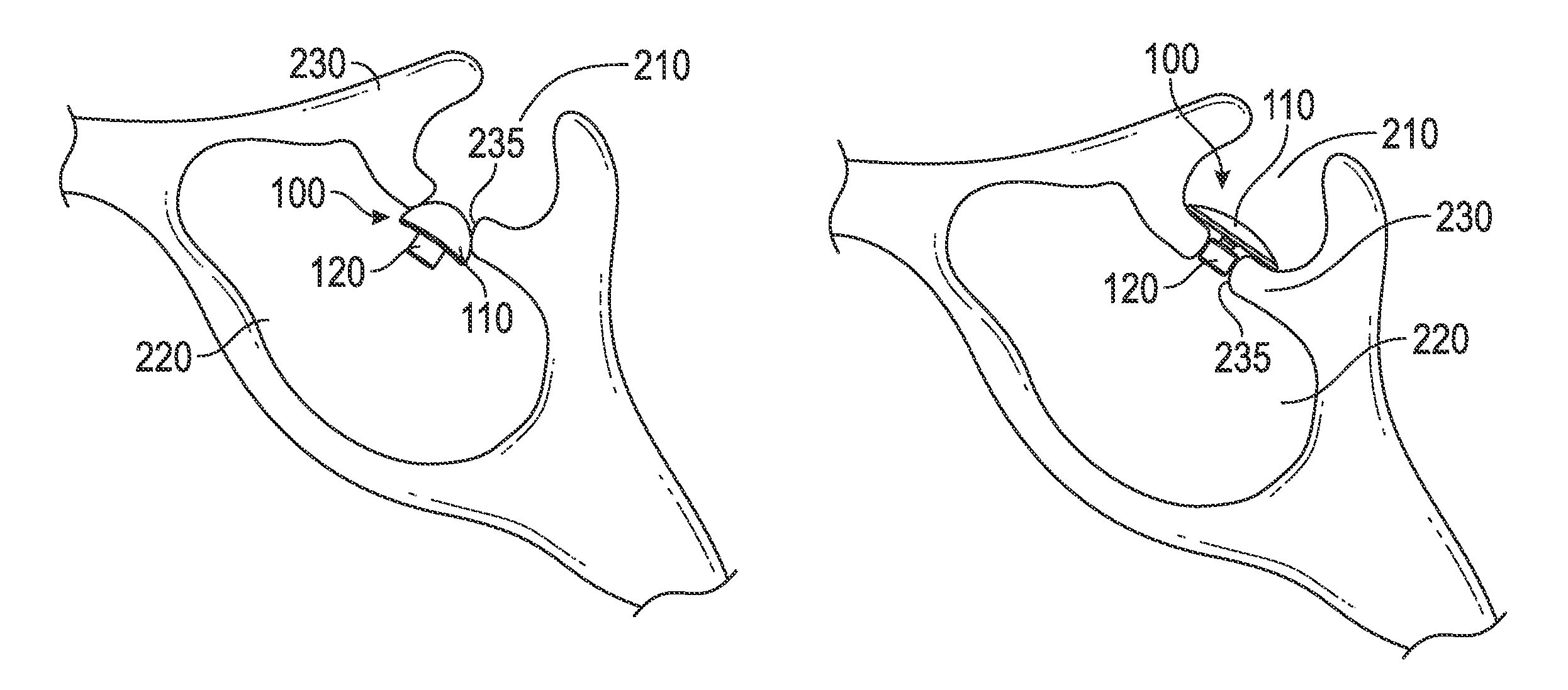
[0019]The device disclosed herein includes a mushroom or conical shaped compressible or deformable biocompatible material soft enough to be atraumatic to bone and surrounding neurovascular structures, yet rigid enough to hold form and provide support after insertion into the sella turcica through a hole smaller than an outer diameter of the contact surface or the maximum width of the device.

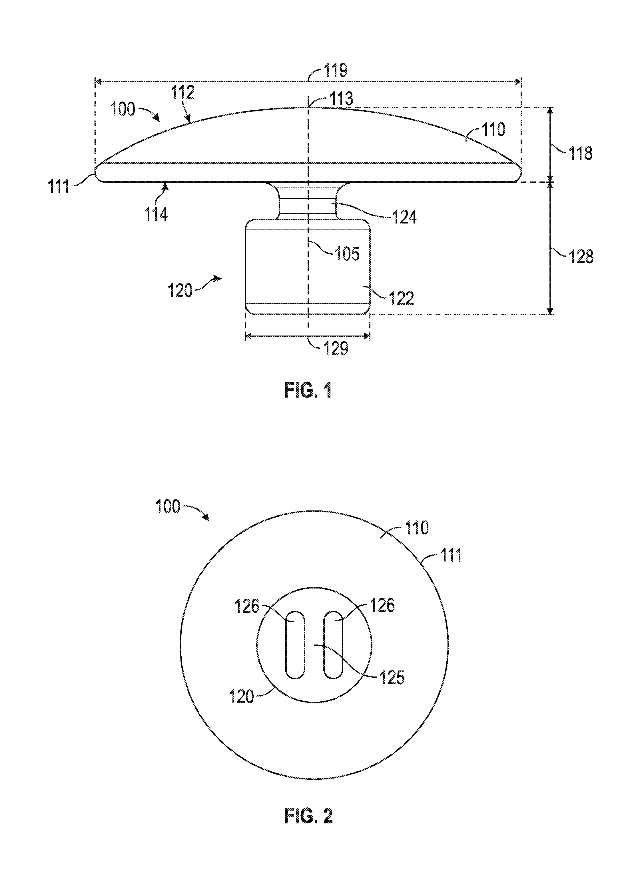
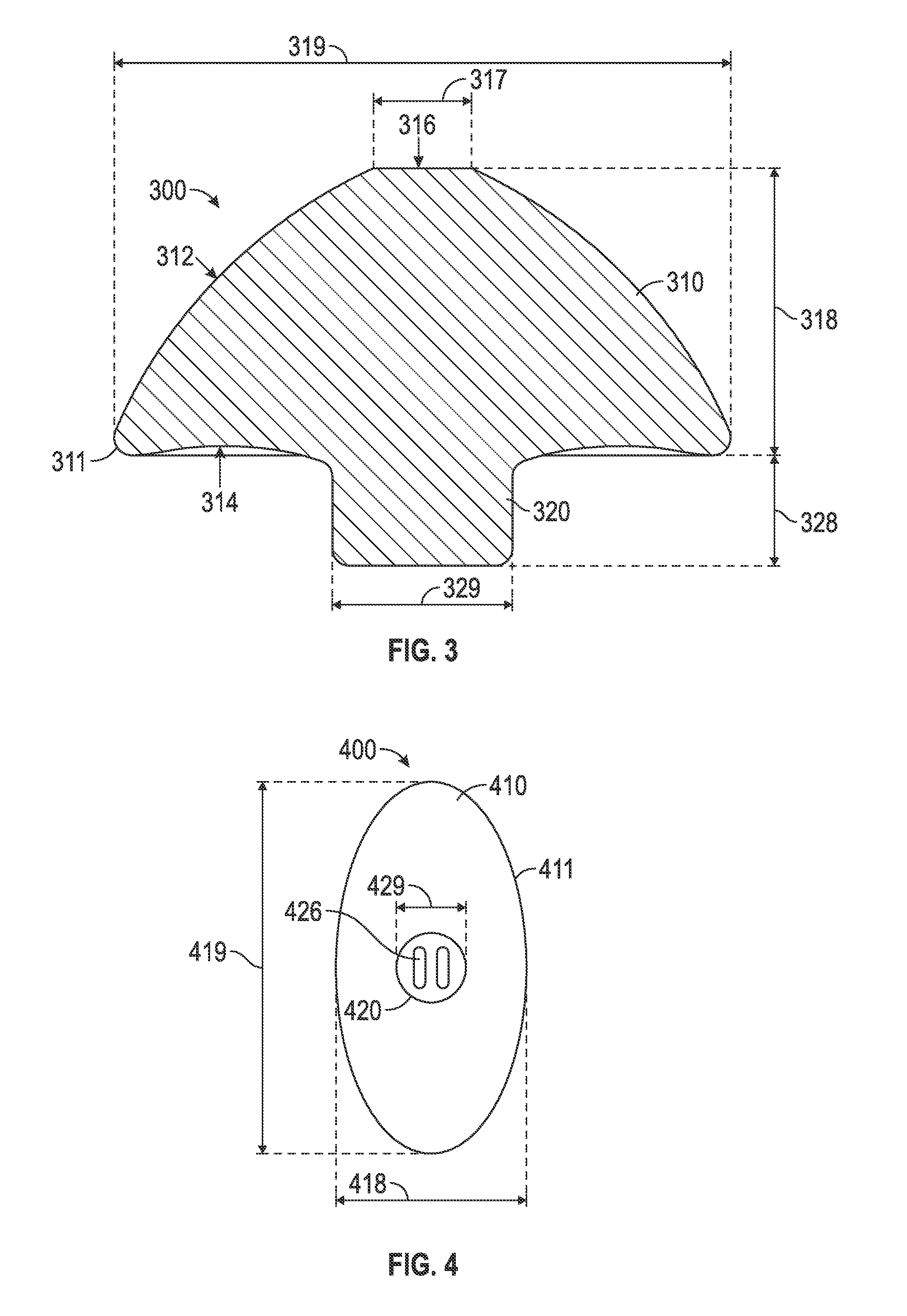
[0020]FIG. 1 is a side view of an exemplary embodiment of a cranial base implant device (“device”) 100. Device 100 includes a cap 110 and a stem 120. Cap 110 may be rounded and may have a spherical, ellipsoidal or conical shape. The ellipsoidal shapes may include oblate and prolate spheroids. In the embodiment shown in FIG. 1, cap 110 is the shape of a spherical cap, a region of a sphere above a given plane; more particularly cap 110 is a spherical cap with a cap height 118 less than the radius of the sphere. In some embodiments, cap 110 is the shape of a hemisphere, a spherical cap with the plan...


PUM


Abstract

A device for closing a hole in a sphenoid bone that acts as a barrier between a sella turcica and a sphenoid sinus is disclosed. The device includes a cap and a stem. The cap is formed of a deformable material. The cap includes a contact surface and an upper surface. The contact surface forms a base of the cap and includes an outer perimeter. The upper surface includes an apex offset from the contact surface. The upper surface extends from the apex to the outer perimeter. The stem extends from the contact surface in a direction opposite the apex.

Description

RELATED APPLICATION[0001]The present application claims the benefit of co-pending U.S. provisional patent application no. 61 / 738,305 filed on Dec. 17, 2012, the contents of which are incorporated herein by reference in their entirety.TECHNICAL FIELD[0002]The present invention relates generally to an implant device used to close a hole or defect in the sphenoid bone that acts as a barrier between the sella turcica and the sphenoid sinus.BACKGROUND[0003]Endoscopic transphenoidal pituitary tumor removal involves removing the floor of the sella turcica, opening the dura mater, and then removing the tumor with a variety of microsurgical techniques. These tumors can extend beyond the sella turcica and intwine with the meninges. Surgical removal of a tumor may disrupt the meninges, leading to the leakage of spinal fluid into the surgical field. Because the spinal fluid is under pressure, the fluid may continue to leak and maintain a communication between the intracranial space and the nasa...

Claims


Application Information

Patent Timeline
IPC IPC(8): A61F2/02A61B17/00A61F2/28A61B19/00
CPCA61B17/0057A61F2/2875A61B2017/00659A61B2019/208A61F2002/2885A61B2090/103
Inventor FITZGERALD, PATRICK J.MCKINNEY, R. TODDBENKE, JASPERMITCHELL, SEAN JEFFREY
Owner FITZGERALD PATRICK J
Why Eureka
Try Eureka