Method for separation and characterization of microorganisms using identifier agents

a technology of identifier agent and microorganism, which is applied in the field of detecting, isolating and/or identifying microorganisms in samples, can solve the problems of bloodstream infection, high morbidity and mortality, and take several days to perform, so as to reduce the risk of handling infectious materials and/or contaminating samples, the effect of characterization and/or identification of microorganisms more quickly and faster

Active Publication Date: 2015-09-08
BIOMERIEUX INC
View PDF45 Cites 10 Cited by
  • Summary
  • Abstract
  • Description
  • Claims
  • Application Information

AI Technical Summary

Benefits of technology

[0014]The present invention provides methods for isolating, characterizing and / or identifying microorganisms in a sample. The methods allow for the characterization and / or identification of microorganisms more quickly than prior techniques, resulting in faster diagnoses (e.g., in a subject having or suspected of having septicemia) and identification of contaminated materials (e.g., foodstuffs and pharmaceuticals). The steps involved in the methods of the invention, from obtaining a sample to characterization and / or identification of microorganisms, can be carried out in a very short time frame to produce clinically relevant actionable information, e.g., in less than about 120 minutes. Additionally, the methods of the invention can be fully automated, thereby reducing the risk of handling infectious materials and / or contaminating samples.
[0030]In one embodiment, the separation is carried out by layering the sample over a density cushion in a container and centrifuging the container to pellet the microorganisms while the sample medium remains on top of the density cushion. In another embodiment, the container has an optical window at the bottom and / or sides so that the microorganism pellet can be interrogated spectroscopically. The microorganisms can be identified by comparing the spectrum of the pellet to a spectrum or spectra of known microorganisms. The ability to identify microorganisms directly in the pellet without further handling enhances the safety of microbial identification.

Problems solved by technology

Bloodstream infections are associated with high morbidity and mortality, yet current diagnostic methods, of culture followed by biochemical identification and antibiotic susceptibility testing, can take several days to perform.
Typically, empiric therapy is initiated based on clinical symptoms, and test results only impact clinical decisions when the initial therapy fails.
Molecular amplification methods have been proposed to fill this need, but serious challenges to this approach remain.
These direct-from-the-bottle tests are not appropriate for all microorganisms (e.g., Gram-positive cocci), are not validated by the test manufacturers, and generally take 3-8 hours to provide results.
Optical spectroscopy methods, such as intrinsic fluorescence (IF), infrared spectroscopy (FTIR), or Raman spectroscopy, and mass spectrometry methods such as MALDI-TOF, have the potential to allow for identification of microorganisms very quickly, but may encounter interference from the many highly fluorescent and absorptive compounds present in liquid microbiological culture media and in clinical samples such as blood or combinations thereof.
However, the '135 application does not discuss identification and quantification of microorganisms from complex biological samples, such as blood.
However, these methods have several drawbacks when attempting to separate and characterize microorganisms from complex samples such as blood-containing culture media.
The resultant microbial preparations often contain contaminating red blood cells, platelets, lipid particles, plasma enzymes and cellular debris, which can cause poor results.
These methods are also very labor-intensive and unsafe due to steps which can result in aerosol exposure of potentially dangerous pathogens to the user.

Method used


Image

  • Method for separation and characterization of microorganisms using identifier agents
  • Method for separation and characterization of microorganisms using identifier agents
  • Method for separation and characterization of microorganisms using identifier agents

Examples


example 1

Rapid Microbial Isolation and Identification Method

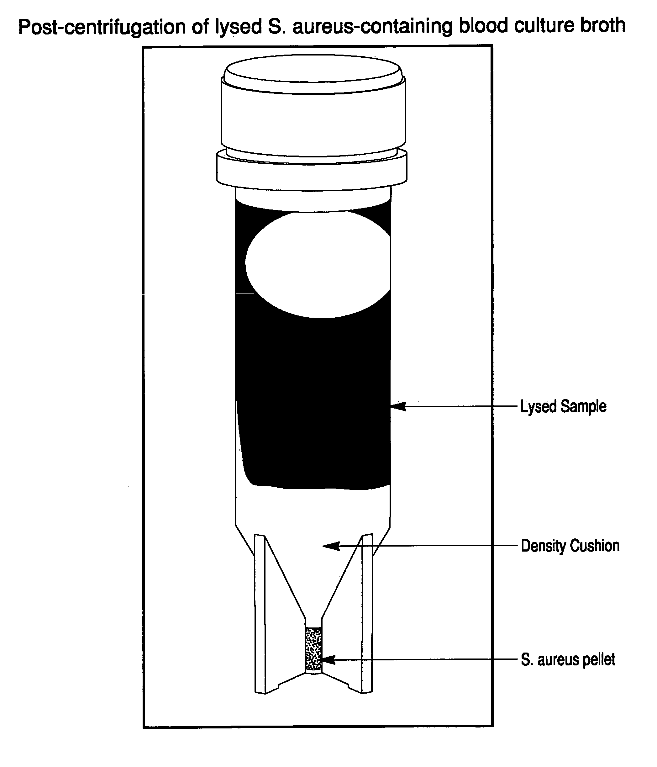
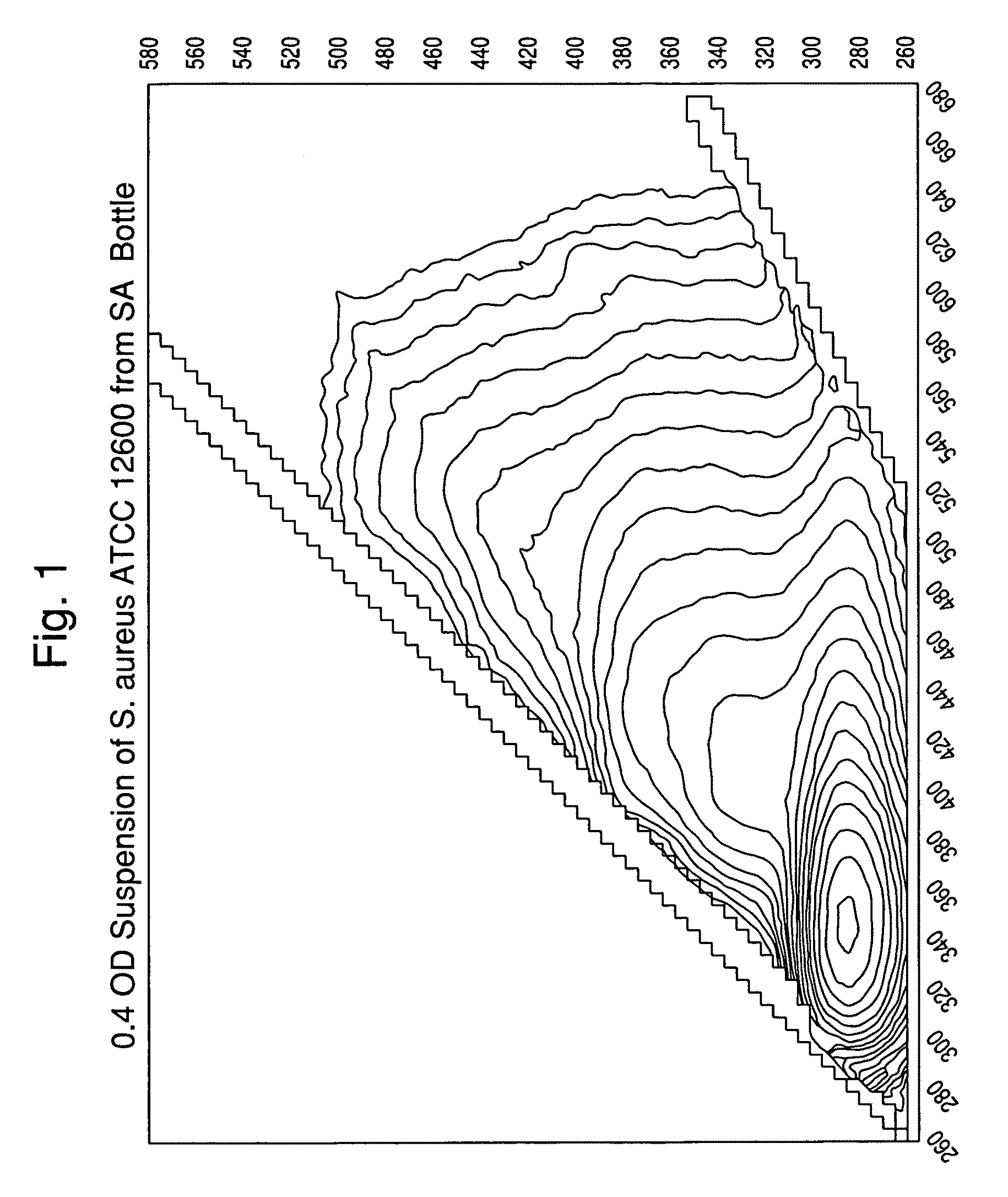
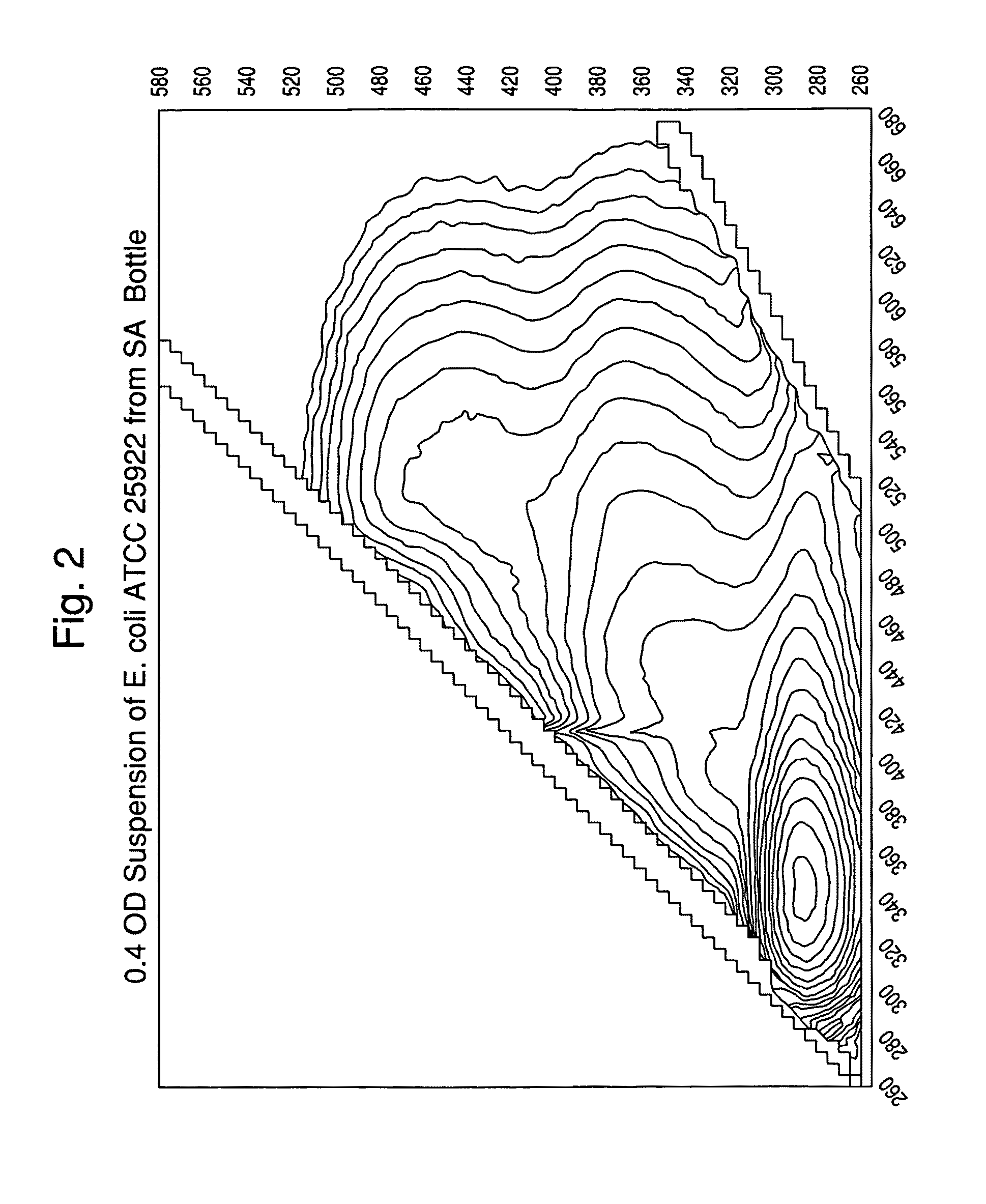
[0119]A suspension of colloidal silica (0.2-0.5 mL; 1.040-1.050 gm / mL density) was added to several conical microcentrifuge tubes. Lysed positive BacT / ALERT® SA blood culture broth samples (0.5-1.0 mL) were overlaid onto the colloidal silica suspension. Alternatively, the colloidal silica solution can be added underneath the lysed blood culture broth using a needle or canula. Positive broth from cultures containing the following microorganisms were tested:[0120]E. coli, ATCC 25922[0121]E. faecalis, ATCC 29212[0122]S. aureus, ATCC 12600[0123]P. aeruginosa, ATCC 10145

[0124]The tubes were capped, and then spun in a microcentrifuge for 2 min at about 10,000 g at room temperature (20-25° C.). The supernatants were aspirated, then the purified microbial pellets were resuspended in 0.45% w / v NaCl to an optical density @ 660 nm of 0.40.

[0125]One portion of each suspension was transferred to an acrylic cuvette and scanned in a spectrofluorim...

example 2

EDTA in Lysis Buffer as an Identifier Agent

[0127]Many lysis buffers, particularly those used for molecular methods, contain chelating agents such as EDTA to assist in the solubilization step. We assessed the impact of adding EDTA to the TX100-CAPS lysis buffer base using a panel of Gram-negative and Gram-positive microorganisms. Table 1 shows the rapid inhibitory effect of EDTA on P. aeruginosa and A. baumanii, but not on two other Gram-negative rods, B. cepacia and K. pneumoniae, or the Gram-positive S. aureus. Note that while EDTA was inhibitory to both P. aeruginosa and A. baumanii, the changes in major intrinsic fluorophores were very different between these two species. P. aeruginosa was the only organism tested that had a significant drop in both NADH and tryptophan fluorescence following EDTA treatment.

[0128]These experiments represents a good example of how certain additives or identifiers can be added to the lysis buffer to rapidly alter the base microbial intrinsic fluores...

example 3

Rapid In Situ Microbial Enzyme Assay

[0130]Several closely-related members of the Enterobacteraciae family can be differentiated by the presence or absence of an enzyme known as pyroglutamyl peptidase. For example, E. aerogenes is positive while E. cloacae is negative; C. freundii is positive and E. coli is negative.

[0131]To test for microbial pyroglutamyl peptidase (Pyr A), positive blood culture broth were treated as follows:[0132]1. A 2.0 mL sample of positive broth was mixed with 1.0 mL of selective lysis buffer (0.45% w / v Brij® 97+0.3 M CAPS, pH 11.7), and then placed in a 37° C. water bath for 1 minute.[0133]2. A 1.0 mL sample of lysate was overlayed onto 0.5 mL of density cushion (24% w / v cesium chloride+10 mM Hepes ph 7.4+0.005% Pluronic F-108) supplemented with 300 ug / mL L-pyroglutamic-acid-7AMC, contained in a custom-built optical separation tube. A polypropylene ball was present on the surface of the density cushion to facilitate loading without disturbing the two aqueous ...


PUM

PropertyMeasurementUnit
temperatureaaaaaaaaaa

Abstract

The present invention is directed to a method for separating, characterizing and / or identifying microorganisms in a test sample. The method of the invention comprises an optional lysis step for lysing non-microorganism cells that may be present in a test sample, followed by a subsequent separation step. The method may be useful for the separation, characterization and / or identification of microorganisms from complex samples such as blood-containing culture media. The invention further provides for the use of one or more identifier agents and interrogating the microorganism sample and / or said one or more identifier agents to produce measurements which characterizing and / or identifying the microorganism based on the produced measurements and / or the presence or absence of the identifier agent or a metabolized form of the identifier agent in the microorganism sample.

Description

CROSS REFERENCE TO RELATED APPLICATION[0001]This application claims the benefit of U.S. Provisional Patent Application No. 61 / 110,187, entitled, “Method and System for Detection and / or Characterization of a Biological Particle in a Sample”, filed Oct. 31, 2008, which is incorporated herein.FIELD OF THE INVENTION[0002]The present invention relates to methods and systems for detecting, isolating and / or identifying microorganisms in a sample. Moreover, the present invention is directed to a method for the enhanced characterization and / or identification of microorganisms using identifier agents.BACKGROUND OF THE INVENTION[0003]The detection of pathogenic microorganisms in biological fluids should be performed in the shortest possible time, in particular in the case of septicemia for which the mortality remains high in spite of the broad range of antibiotics which are available to doctors. The presence of biologically active agents such as a microorganism in a patient's body fluid, espec...

Claims


Application Information

Patent Timeline
Patent Type & Authority Patents(United States)
IPC IPC(8): C12Q1/04G01N21/65G01N33/68G01N21/35G01N21/3581G01N21/47G01N21/64
CPCG01N21/65C12Q1/04G01N33/6848G01N21/3581G01N21/47G01N21/64G01N21/35C12M1/34C12Q1/24C12Q1/37C12Q1/6816
Inventor WALSH, JOHNHYMAN, JONES M.THORPE, THURMANCLAY, BRADFORD
Owner BIOMERIEUX INC
Why Eureka
Try Eureka