Bin1 as a prognostic marker in cardiovascular disease

a prognostic marker and cardiovascular disease technology, applied in the field of cardiovascular disease, can solve the problems of cardiac mortality, poor outcome, 2000 hearts available, etc., and achieve the effect of increasing the risk of poor outcom

Active Publication Date: 2015-10-06
RGT UNIV OF CALIFORNIA
View PDF11 Cites 5 Cited by
  • Summary
  • Abstract
  • Description
  • Claims
  • Application Information

AI Technical Summary

Benefits of technology

The present patent provides methods for using BIN1 expression levels to evaluate the risk of a poor outcome in patients diagnosed with congestive heart failure. This can be applied to patients who are heart transplant candidates, as well as those who are undergoing treatment or are in an immunosuppression therapy. The method involves measuring BIN1 expression levels in heart tissue obtained from the patient and using them to determine the risk of a poor outcome. Patients with a decreased BIN1 expression level or an increased IDF ratio are at increased risk of a poor outcome. This can help healthcare professionals better understand the severity of the patient's condition and decide on appropriate treatment options.

Problems solved by technology

However, only 2000 hearts are available each year for heart transplant.
Poor outcome can include, for example, cardiac mortality, the need for mechanical device support (e.g., a left ventricular assist device (LVAD) implant), or a left ventricular ejection fraction of less than 25%.
In certain embodiments, a decreased BIN1 expression level compared to a reference BIN expression level is positively correlated to an increase in the risk of a poor outcome.
In certain embodiments, the IDF ratio of the patient is compared to a reference IDF and an IDF ratio greater than than the reference IDF ratio is indicative of an increased risk of a poor outcome

Method used


Image

  • Bin1 as a prognostic marker in cardiovascular disease
  • Bin1 as a prognostic marker in cardiovascular disease
  • Bin1 as a prognostic marker in cardiovascular disease

Examples


example 1

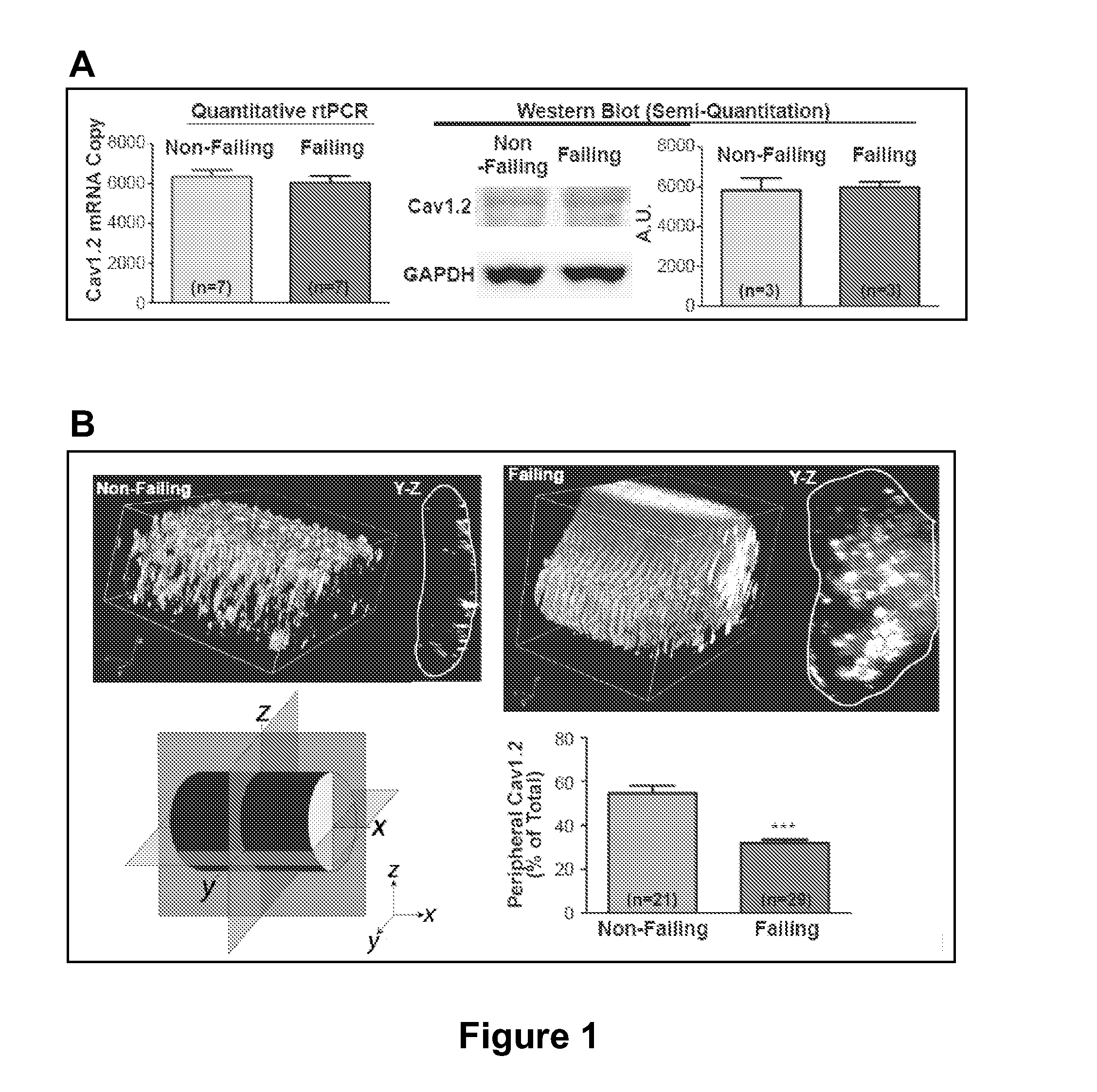
CaV1.2 Has an Intracellular Distribution in Failing Heart

[0148]Cardiac excitation-contraction (EC) coupling, a process essential to each heartbeat, links electrical excitation of the myocyte to its mechanical contraction. EC coupling begins with local calcium entry through CaV1.2 channels that then causes a large release of calcium by the intracellular sarcoplasmic reticulum (Fabiato, 1983). Ryanodine receptors on the sarcoplasmic reticulum (SR) are the local calcium sensors and release channels (Cheng et al., 1993; Inui et al., 1987; Pessah et al., 1985). In ventricular cardiomyocytes, close association of CaV1.2 channels and ryanodine receptors is necessary for efficient calcium-induced-calcium release (CICR) (Bers, 2002). Synchronous CICR occurs at the EC-couplons, where 10-25 L-type calcium channels and 100-200 ryanodine receptors coincide and cooperate with each other (Bers, 2001). In order to locally approximate the SR bound ryanodine receptors, CaV1.2 channels are necessarily...

example 2

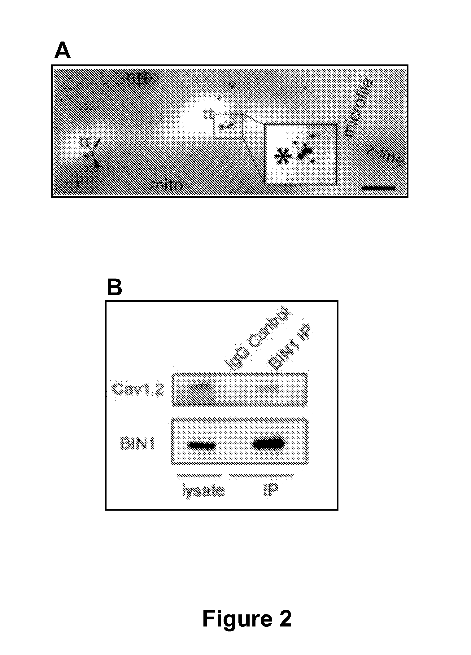
BIN1 Targets CaV1.2 to T-Tubules

[0152]The mechanism of CaV1.2 targeting to T-tubule membranes was explored. Co-staining of BIN1 and CaV1.2 in both isolated non-failing human cardiomyocytes and mouse cardiomyocytes indicates that BIN1 outlines T-tubules and CaV1.2 is enriched along such invaginations and colocalizes with BIN1. For higher resolution imaging, transmission electron microscopy with dual immunogold labeling was used to identify CaV1.2 and BIN1 on T-tubule ultrastructures in adult mouse cardiomyocytes. A representative image in FIG. 2A indicates that BIN1 (small 10 nm dots) and CaV1.2 (large 15 nm dots) are enriched and cluster within 10-50 nm of each other, occurring at T-tubular membrane structures.

[0153]Given that CaV1.2 is closely associated with the tubulogenesis protein BIN1 at T-tubules, it is possible that BIN1 specifically attracts delivery of CaV1.2 to T-tubules. This possibility was tested using the reductionist approach of studying the association between BIN1 ...

example 3

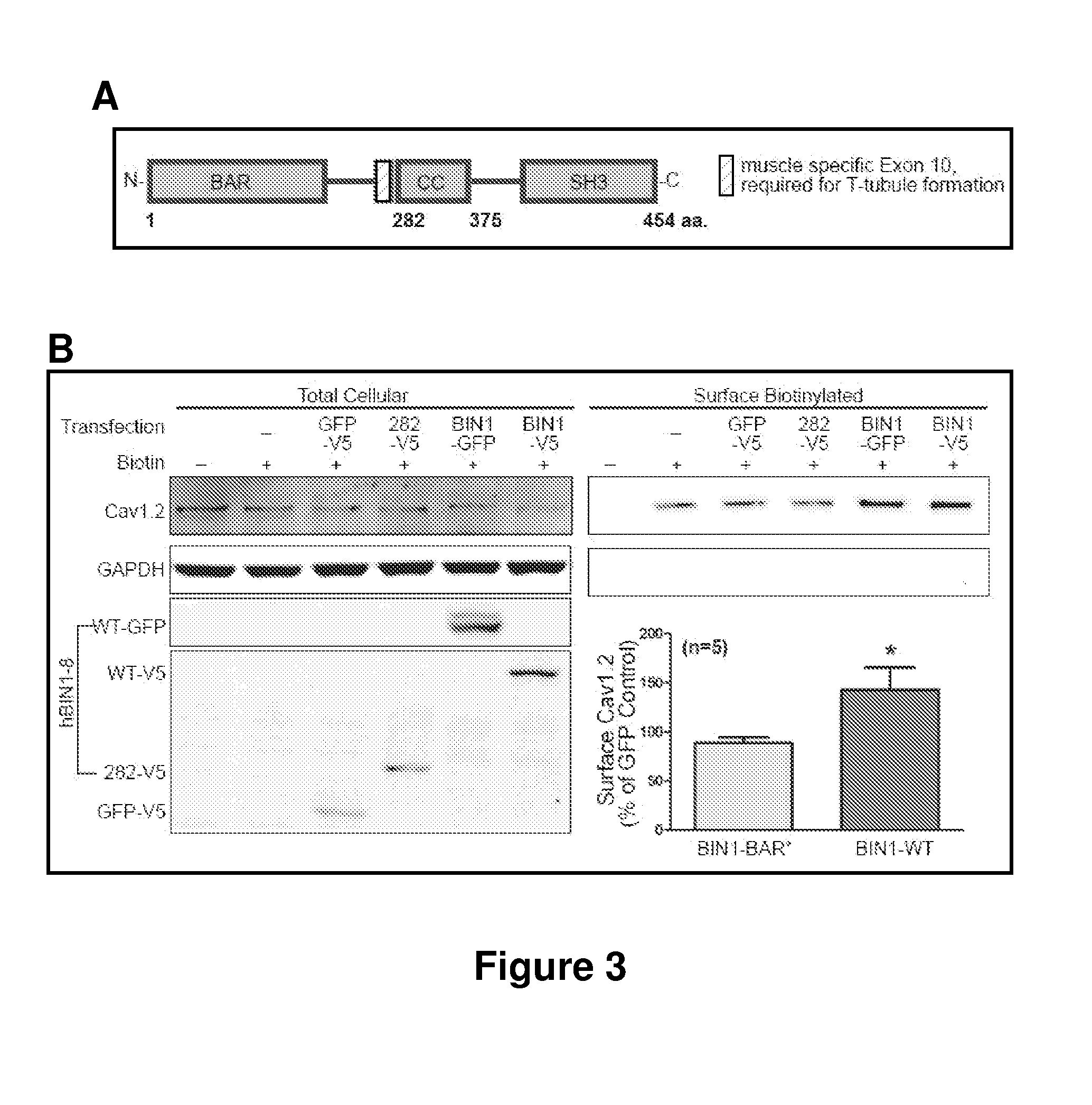
CaV1.2 is Targeted to BIN1, not T-Tubules

[0155]It was determined whether it is BIN1 protein or T-tubule structures that is sufficient for CaV1.2 targeting. Wild-type BIN1 (BIN1-WT, 1-454aa) has an N-terminal BAR domain followed by 15 amino acids (aa) encoded by exon 10 which is required for phospholipid binding and T-tubule formation, a coil-coiled linkage domain, and a C-terminal SH3 domain for protein-protein interaction (FIG. 3A) (Lee et al., 2002; Nicot et al., 2007). A previously published C-terminal truncated BIN1-BAR* (1-282aa, BAR+Exon10), which retains the ability to induce membrane invagination was created (Lee et al., 2002). BIN1-BAR* cannot attract endogenous CaV1.2 to the nascent T-tubule structures such as those in HL-1 cells. In an HL-1 cell transfected with BIN1-WT, endogenous CaV1.2 is distributed along BIN1 structures. In contrast, in cells transfected with BIN1-BAR*, CaV1.2 shows poor colocalization with the BIN1 structures. The effect of BIN1-WT and BIN1-BAR* on ...


PUM


Abstract

The present disclosure provides methods involving use of BIN1 expression levels, in heart tissue, in evaluating the risk of a poor outcome in a patient diagnosed with congestive heart failure. The methods finds use in evaluating patients who are heart transplant candidates as well as in assessing therapy options and efficacy of treatment in congestive heart failure patients.

Description

CROSS-REFERENCE TO RELATED APPLICATIONS[0001]This application claims priority benefit to U.S. provisional application Ser. No. 61 / 172,608 filed on Apr. 24, 2009 which is hereby incorporated by reference in its entirety.STATEMENT REGARDING FEDERALLY SPONSORED RESEARCH[0002]This invention was made with government support under Grant No. HL094414 awarded by the NHLBI, National Institutes of Health. The government has certain rights in the invention.TECHNICAL FIELD[0003]This invention relates to cardiovascular disease and methods to assess the same.INTRODUCTION[0004]During the last decade, congestive heart failure (CHF) has burgeoned into the most prominent public health problem in cardiovascular medicine. CHF affects close to 20 million people worldwide and approximately 5 million Americans. In the United States alone, about 400,000 new cases of CHF are diagnosed annually and the condition is responsible for approximately 200,000 deaths per year. About a 100,000 patients diagnosed with...

Claims


Application Information

Patent Timeline
Patent Type & Authority Patents(United States)
IPC IPC(8): G01N33/566C12Q1/68C07K14/47G01N33/68
CPCC12Q1/6883C07K14/4716C07K14/4747G01N33/6893G01N2333/4703G01N2800/325G01N2800/56
Inventor SHAW, ROBINHONG, TING-TINGSMYTH, JAMES
Owner RGT UNIV OF CALIFORNIA
Why Eureka
Try Eureka