Technique for tomographic image recording

a technology of tomographic image and recording, which is applied in the field of tomographic image recording apparatus, can solve the problems of destroying specimens, changing specimens in uncontrolled manners, and reducing radiation intensity with comparable image results, and achieves the effect of larger numerical aperture and higher resolution

Active Publication Date: 2016-02-02
LASER ZENT HANNOVER
View PDF18 Cites 18 Cited by
  • Summary
  • Abstract
  • Description
  • Claims
  • Application Information

AI Technical Summary

Benefits of technology

[0016]The apparatus can acquire the scattered radiation escaping by reason of an individual pencil beam (at right angles to the beam direction). In comparison with conventional techniques, in this way a considerably larger proportion of the scattered radiation is capable of being acquired by the detector unit. Accordingly, the signal-to-noise ratio of the acquisition can be significantly improved. Also, a radiation intensity (with identical or improved image quality) introduced by the pencil beam can be diminished. In addition, a signal mixing of pencil beams that have been offset (parallel) relative to one another (i.e. various measuring positions) can be excluded. Also, the specimen volume can be acquired with a uniform sensitivity, insofar as the same detector unit acquires all the measuring positions. In particular, ring artefacts (such as frequently arise in the course of the tomographic reconstruction of a spatially resolved acquisition) can be avoided. In comparison with the spatially resolved acquisition of conventional techniques, the acquisition by the detector unit may be regarded as “zero-dimensional”. Furthermore, a more homogenous (in effect) illumination of the specimen volume, in particular at right angles to the beam direction, in comparison with a simultaneous illumination (which, for instance, employs a diffuser) can be obtained. Advantageously, for an excitation of the scattered radiation that is as homogeneous as possible the light-source comprises a laser that is stable (over an image acquisition time) by way of radiation-source.
[0027]The light-source may furthermore have been designed to generate the pencil beam in the visible spectrum, in the ultraviolet spectrum, or in the infrared spectrum. Generally, the pencil beam may lie within the electromagnetic spectrum having (vacuum) wavelengths from 300 nm to 2000 nm. This spectrum is suitable for non-destructive acquisition of an image of a specimen of deoxyribonucleic acid (DNA). A preferred spectral range of the pencil beam encompasses 323 nm to 750 nm or 355 nm to 850 nm (in the case of fluorescent radiation by way of scattered radiation). In conjunction with absorption tomography (by means of a transmission photo-detector), the pencil beam may (additionally) exhibit radiation from 200 nm to 323 nm (or from 200 nm to 355 nm). Furthermore, the pencil beam may excite the scattered radiation at least partly in the extreme-ultraviolet spectrum (XUV) or in the far-infrared spectrum (FIR). In the case of a water-free specimen, the pencil beam may (additionally) exhibit radiation within the range above 2000 nm. With a view to multi-photon excitation of the fluorescent radiation (in short: “multi-photon fluorescence”), each wavelength specification above (of a spectral range boundary) can be multiplied by a factor of 2 (in the case of a two-photon excitation), by a factor of 3 (in the case of a three-photon excitation), etc. With a view to multi-photon excitation, the light-source may emit brief intense pulses. A femtosecond laser or a (Q-switched) laser with pulse durations of a few picoseconds can be employed as radiation-source for multi-photon excitation. The multi-photon fluorescence can enable the acquisition of a greater depth (in the beam direction).
[0032]A first collimator of the scattered-radiation collector may include a strongly refracting lens or a condenser lens which has been designed to collimate the acquired scattered radiation. Alternatively or additionally, the scattered-radiation collector may exhibit as first collimator a tapered light guide (tapered light pipe, TLP) which has been designed to collimate the acquired scattered radiation. The light guide may be a reflection-coated (on the inside) optical hollow-core conductor. Alternatively, the light guide may be a solid glass rod, so that the scattered radiation is guided by total reflection (for instance, to the air surrounding the solid glass rod). Alternatively or in supplement, the solid glass rod may have been vapour-coated with a dichroic layer. The dichroic layer can reflect the scattered radiation for steep angles of incidence (i.e. small angles of incidence relative to the normal to the inner surface of the optical hollow-core conductor). This can enable reflection (on the dichroic layer) to occur for steep angles, and total reflection to occur for shallow angles (i.e. for large angles of incidence with the normal to the inner surface of the optical hollow-core conductor).
[0048]The light-source may furthermore exhibit a focusing lens system which has been arranged between the optical control element and the specimen holder. The focusing lens system may comprise a focusing lens or a focusing-lens system for forming the pencil beam (with the defined beam within the specimen holder). Preferentially, the focusing lens system exhibits a telescopic lens system and a focusing lens. To the advantage of a higher resolution, the focusing lens system can widen the beam (downstream of the rasterisation mirrors). In this way, a width of the rasterisation mirrors may be smaller than the beam width. The focusing lens system can radiate the pencil beam into the specimen holder with a larger numerical aperture. In this way, an incident numerical aperture of NAE=0.2 is attainable. (The incident numerical aperture, NAE, is not to be confused with the acquired numerical aperture, NA.) A first focal plane of the focusing lens system may include (substantially) a position of the rasterisation mirror. A second focal plane of the focusing lens system may include (substantially) a position within the specimen holder, preferentially a position of the beam waist or of the axis of rotation within the specimen holder.

Problems solved by technology

The known techniques for spatial image acquisition can, by reason of a radiation intensity acting on the specimen, distort the image acquisition, change the specimen in uncontrolled manner or, particularly in the case of in vivo microscopy, destroy the specimen.
Also, the technical effort in order with the prior techniques to diminish the radiation intensity with comparable image results may rise considerably.

Method used


Image

  • Technique for tomographic image recording
  • Technique for tomographic image recording
  • Technique for tomographic image recording

Examples


third embodiment

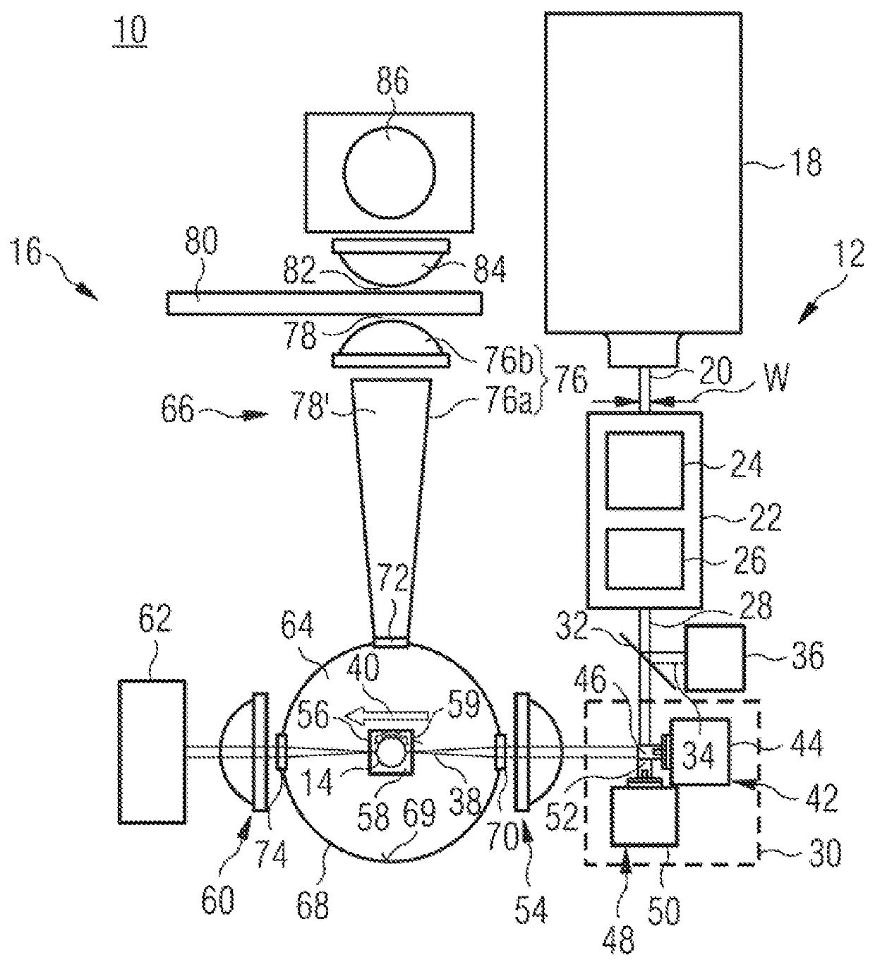
[0069]The specimen holder 14 has been rotatably arranged in a specimen vessel 56, preferentially a cell. A liquid (which in the case of the specimen vessel 14 coincides, at least with regard to its refractive index, with the aforementioned liquid) adapted to the specimen as regards its refractive index is located between the specimen holder 14 and the specimen vessel 56. The rotation relates to a relative rotation between, on the one hand, the specimen holder 14 and, on the other hand, at least the light-source 12 and the specimen vessel 56. In principle, a “boundary” or “interface” of the rotation extends between the specimen holder 14 and the specimen vessel 56 (particularly since the specimen vessel 56 may exhibit entrance windows and exit windows, described below, for the pencil beam 38, the location of which, with a view to simplified reconstruction, should not be rotated in relation to the pencil beam 38). An axis of rotation of the rotation is vertical (perpendicular to the p...

fourth embodiment

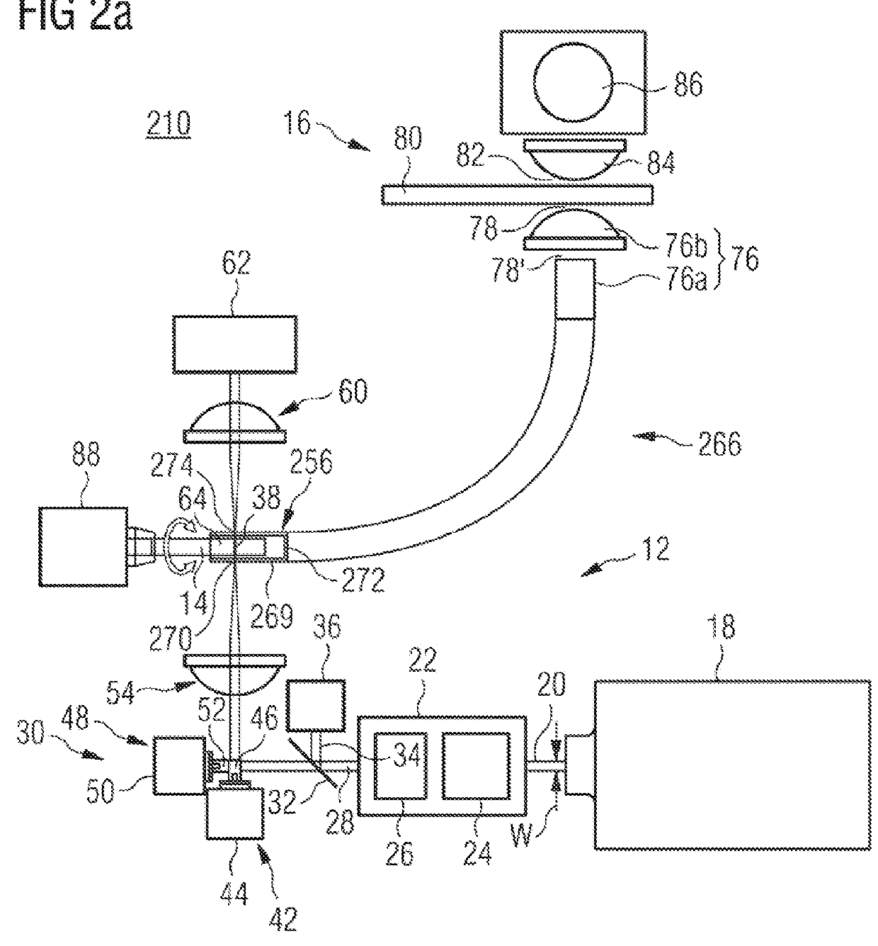
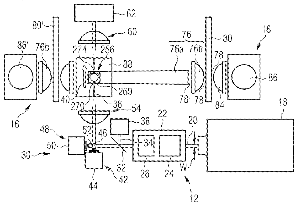
[0107]With a view to radiating the pencil beam 38, and optionally for transmission detection, the specimen vessel 256 exhibits an illumination aperture 270 and a transmission aperture 274, respectively. The illumination aperture 270 and, where appropriate, the transmission aperture 274 correspond (functionally) to aperture 70 and to aperture 74, respectively. As described above in connection with apertures 70, 72, 74, with a view to minimising radiation losses apertures 270 and 274 take the form of narrow slits in the x-direction, and the optical control element 30 rasterises the pencil beam 38 only in the x-direction. For measuring positions with y-offset, the specimen holder 14 can be moved relative to all other components in the y-direction, whereby the specimen holder 14 or the other components are stationary, as has been described above for the first exemplary embodiment. The reflecting surface 269 exhibits opposing beam-transparent slits by way of apertures 270 and 274. In the...

fifth embodiment

[0109]the specimen vessel 256 shown in FIG. 2 for fluorescent radiation by way of scattered radiation includes a transparent base body which exhibits a dichroic layer on the side surfaces. Alternatively or in supplement, the side surfaces of the specimen vessel consist of a dichroic filter or an interference filter. In this way, the side surfaces of the specimen vessel 256 have been designed to transmit the exciting radiation (of the pencil beam 38) and to reflect the fluorescent radiation. In this way, a proportion of the scattered radiation 64 of about 50% or more can be acquired. With regard to the fluorescent radiation (i.e. the scattered radiation 64), the side faces act as reflecting surface 269. Since the side faces are transparent with regard to the exciting radiation (of the pencil beam 38), the side surfaces in addition enable the entrance and exit of the pencil beam 38. That is to say, the entire side surfaces are available by way of illumination aperture 270 or transmiss...


PUM

PropertyMeasurementUnit
wavelengthsaaaaaaaaaa
lengthaaaaaaaaaa
sizeaaaaaaaaaa

Abstract

The invention relates to a device (10; 210; 310; 410; 510) for tomographic image recording. The device comprises a sample retainer (14), a light source (12), and a detector unit (16). The light source (12) is designed to produce a pencil beam (38), which has a beam direction (40) and which passes through a sample volume of the sample retainer (14) provided in order to accommodate a sample, and has an optical control element (30), which is able to move the pencil beam (38) passing through the sample retainer (14) transversely to the beam direction (40) while the beam direction (40) remains substantially unchanged. The detector unit (16) is designed to detect at least a portion of scattered radiation (64; 64′) escaping from a section of the pencil beam (38) within the sample volume or the sample retainer (14) in a non-spatially-resolved manner.

Description

CROSS-REFERENCE TO RELATED APPLICATIONS[0001]This application is a National Stage application of PCT International Application Serial No. PCT / EP2011 / 073077, filed Dec. 16, 2011, which claims priority from German Application No. 10 2010 063 412.3, filed Dec. 17, 2010.TECHNICAL FIELD[0002]The invention relates generally to a technique for acquiring a sectional image or a three-dimensional image. In particular, the invention relates to an apparatus for tomographic acquisition of an image of a spatial object.TECHNICAL BACKGROUND[0003]Resolution and contrast of existing techniques for spatial image acquisition, particularly for microscopy of living organisms, have been significantly improved in recent years. Widespread techniques with high resolution, such as the confocal microscopy described in printed publication DE 43 26 473, are based on the spatially resolved acquisition of an overlap of illumination volume and acquisition volume. The volume of the overlap determines the resolution....

Claims


Application Information

Patent Timeline
Patent Type & Authority Patents(United States)
IPC IPC(8): G01N21/00G01B9/02G01N21/47G01N21/64G01N21/17
CPCG01B9/02091G01N21/4795G01N21/6408G01N21/6458G01N2021/1787G01N2201/065
Inventor LORBEER, RAOUL-AMADEUSMEYER, HEIKOHEIDRICH, MARKOHEISTERKAMP, ALEXANDER
Owner LASER ZENT HANNOVER
Why Eureka
Try Eureka