Oncostatin M (OSM) antagonists for preventing cancer metastasis and IL-6 related disorders
a technology of oncosm and osm, which is applied in the field of cancer treatment, can solve the problems of malignant phenotype formation of putative cancer stem cells, and achieve the effect of reducing tumor cell detachment, proliferation and/or metastasis
- Summary
- Abstract
- Description
- Claims
- Application Information
AI Technical Summary
Benefits of technology
Problems solved by technology
Method used
Image
Examples
example 1
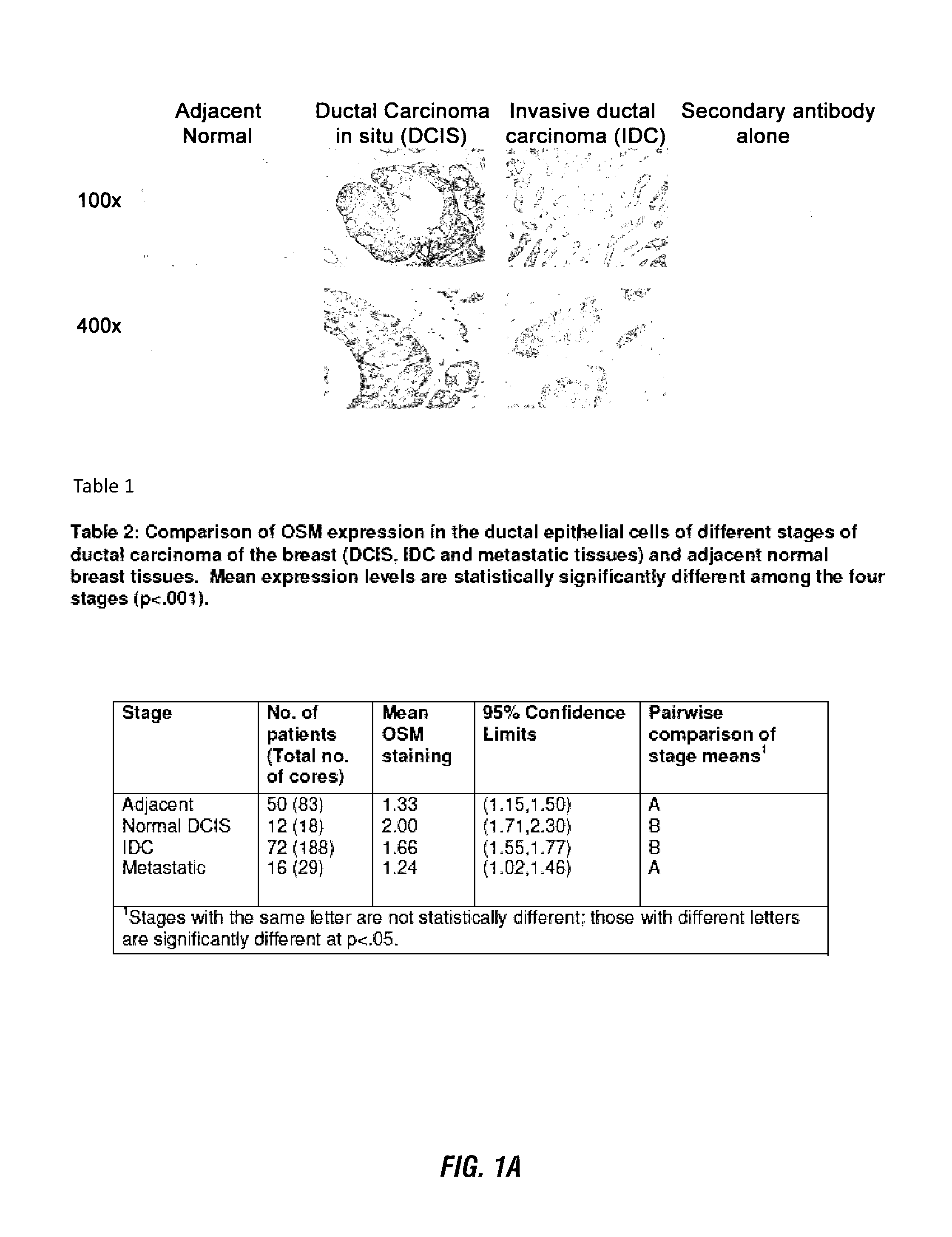
Oncostatin M is Highly Expressed in Early Stages of Ductal Carcinoma of the Breast
[0215]The expression of OSM in a series of TMA breast samples was analyzed by immunohistochemistry (IHC). Treatment of OSM antibody with 10-fold excess OSM blocking peptide followed by IHC resulted in no positive staining, indicating that the antibody is specific to OSM (data not shown).
[0216]Three TMAs containing samples from a total of 72 patients were employed for this study. Of these, 54 patients were from the non-metastatic group and 18 were from the metastatic group. The metastatic group contained samples from patients diagnosed with metastasis of IDC to lymph nodes. All patients in this study were diagnosed with IDC. A total of 12 patients also had DCIS. In addition, the TMAs contained adjacent normal tissues from a total of 50 patients (Table 1).
[0217]Table 1 shows how the 72 patients provided data for the statistical analysis of OSM expression. All of the 72 samples with IDC expressed OSM whil...
example 2
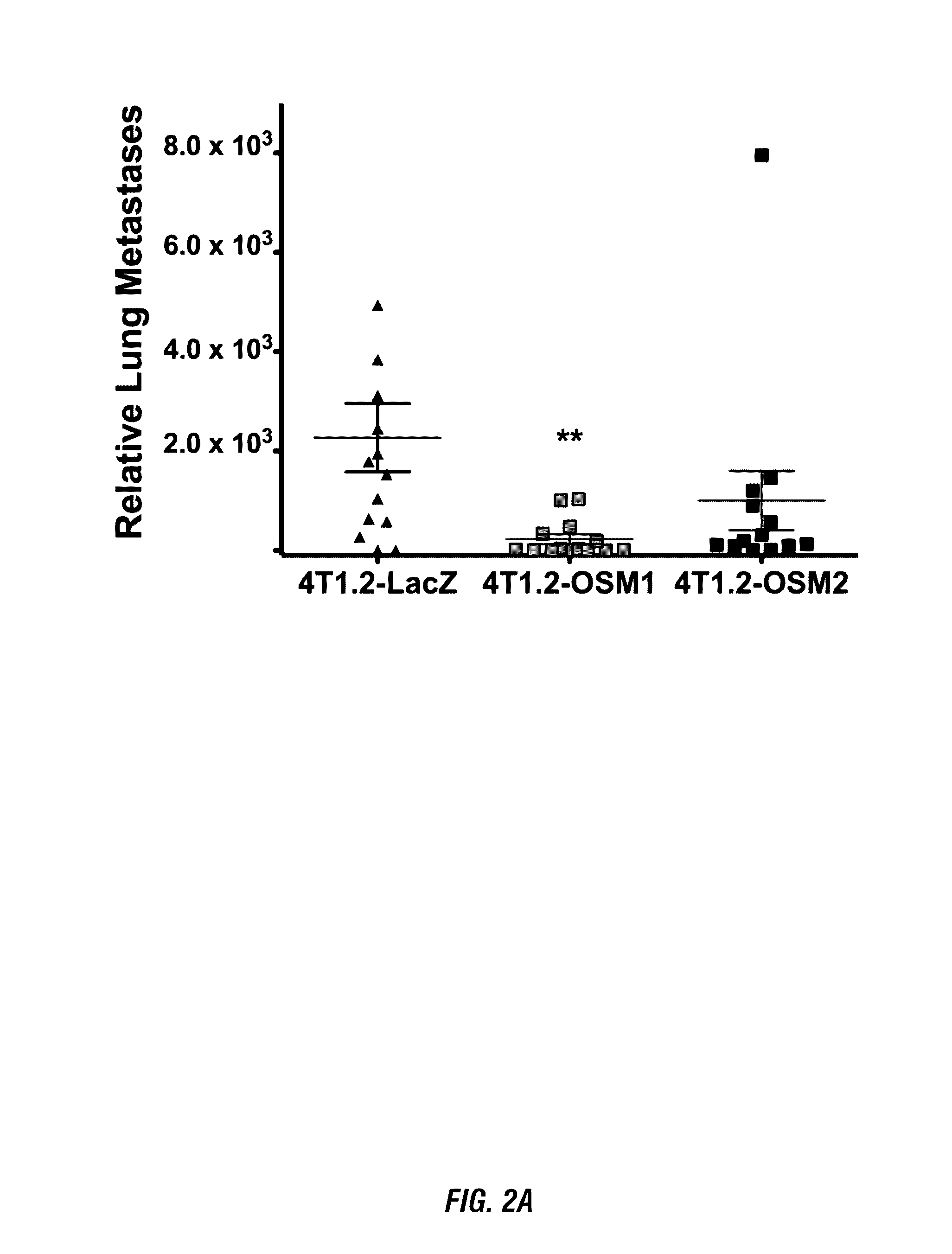
OSM Promotes Mammary Tumor Metastasis to Lung
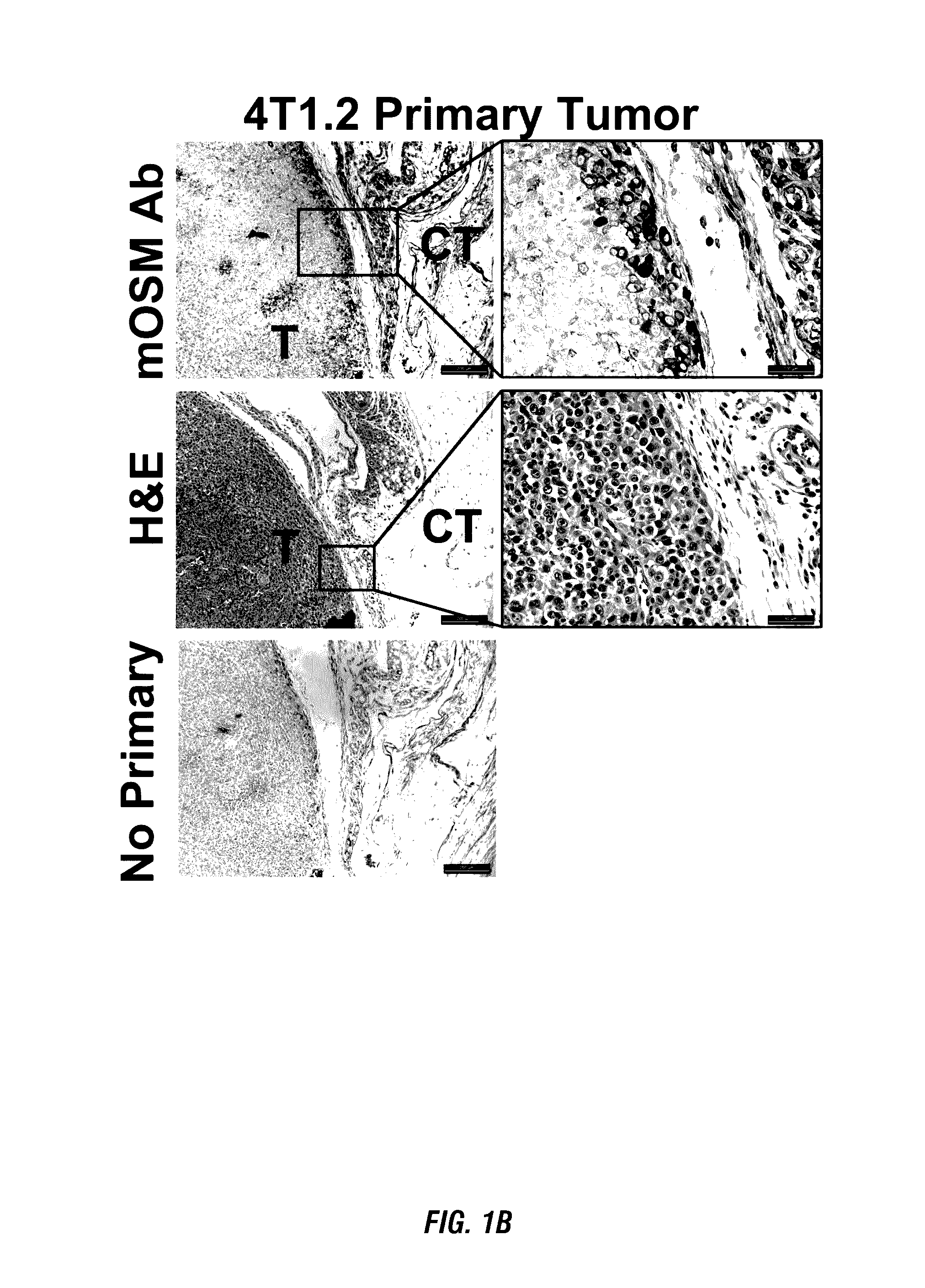
[0219]To examine whether OSM is required for mammary tumor metastasis to lung, we undertook stable knockdown of OSM expression. Two independent OSM shRNA sequences were cloned into the pSilencer4.1 vector and transfected into 4T1.2 mouse mammary tumor cells (T1.2-shOSM1 and 4T1.2-shOSM2) using a procedure known to result in a 3 to 12-fold reduction in OSM expression. To test the effects of OSM on mammary tumor metastasis in vivo, control 4T1.2-LacZ, 4T1.2-shOSM1, and 4T1.2-shOSM2 cells were injected orthotopically into the mammary fat pads of Balb / c mice. Low levels of secreted OSM in the 4T1.2-shOSM2 cells were shown by our lab to result in increased primary tumor growth. However, injection of 4T1.2-shOSM1 cells, which displayed a modest decrease in tumor cell-secreted OSM, did not affect tumor growth in vivo.
[0220]Metastasis to lung in mice injected with control 4T1.2-LacZ, 4T1.2-shOSM1, and 4T1.2-shOSM2 cells were quantified by qPCR. M...
example 3
OSM Expression Increases the Number and Volume of Lung Metastases In Vivo
[0221]To more specifically characterize and quantify the progression of lung metastases seen in vivo after injection of parental 4T1.2, control 4T1.2-LacZ, and 4T1.2-shOSM2 cells, in vivo magnetic resonance imaging (MRI) experiments were performed. Respiratory-gated spin-echo images, with coronal orientation, were collected with sufficient slices (e.g., 21 slices, 0.5 mm thickness) to completely cover the lungs of each animal. Mice were imaged at early (days 20-21), mid (days 25-26), and late (days 29-30) stages of in vivo metastasis (FIG. 3A). For all three cell types, MRI spectra showed essentially no detectable metastasis at the early stages. At mid and late stages, however, readily identifiable metastases were observed in lung images. Lung tumors were manually segmented with IMAGE J (rsbweb.nih.gov / ij), and the number and volume of all metastatic tumors were measured and recorded, on an animal-by-animal bas...
PUM
| Property | Measurement | Unit |
|---|---|---|
| concentration | aaaaa | aaaaa |
| dissociation equilibrium constant | aaaaa | aaaaa |
| dissociation equilibrium constant | aaaaa | aaaaa |
Abstract
Description
Claims
Application Information
 - R&D
- Intellectual Property
- Life Sciences
- Materials
- Tech Scout
- Unparalleled Data Quality
- Higher Quality Content
- 60% Fewer Hallucinations
Browse by: Latest US Patents, China's latest patents, Technical Efficacy Thesaurus, Application Domain, Technology Topic, Popular Technical Reports.
© 2025 PatSnap. All rights reserved.Legal|Privacy policy|Modern Slavery Act Transparency Statement|Sitemap|About US| Contact US: help@patsnap.com


