Method and systems for weight adjustment of an automated breast ultrasound system

an automated breast ultrasound and system technology, applied in the field of medical imaging, can solve the problems of increasing the early breast cancer detection rate, reducing the pressure applied to the scanning apparatus, and fatigue of users, so as to maintain the moveability increase the effective weight of the scanning assembly, and relieve the user's applied force

Active Publication Date: 2017-03-21
GENERAL ELECTRIC CO
View PDF14 Cites 2 Cited by
  • Summary
  • Abstract
  • Description
  • Claims
  • Application Information

AI Technical Summary

Benefits of technology

[0005]In this way, the counterweight and weight adjustment system may counterbalance the scanning assembly while the scanning assembly is positioned on a tissue to be imaged, allowing the scanning assembly to be neutrally buoyant in space during the positioning. Once the scanning assembly has been positioned on the tissue, the effective weight of the scanning assembly may be increased by the weight adjustment system to apply pressure on the tissue, relieving the user's applied force, yet still compressing the tissue for imaging and still maintaining the moveability of the scanning assembly relative to the patient. By doing so, a more consistent amount of pressure may be automatically provided by the scanning assembly rather than relying on the user to apply pressure during the scanning procedure. Further, the patient is not locked in place by the scanning assembly. Thus, high-quality images may be obtained throughout the entirety of the scanning procedure while improving both patient and user comfort.

Problems solved by technology

Thus, the use of volumetric ultrasound scanning in conjunction with conventional x-ray mammography may increase the early breast cancer detection rate.
However, the user may become fatigued and decrease the pressure applied to the scanning apparatus as the scanning progresses, compromising the quality of the acquired images.

Method used


Image

  • Method and systems for weight adjustment of an automated breast ultrasound system
  • Method and systems for weight adjustment of an automated breast ultrasound system
  • Method and systems for weight adjustment of an automated breast ultrasound system

Examples


Embodiment Construction

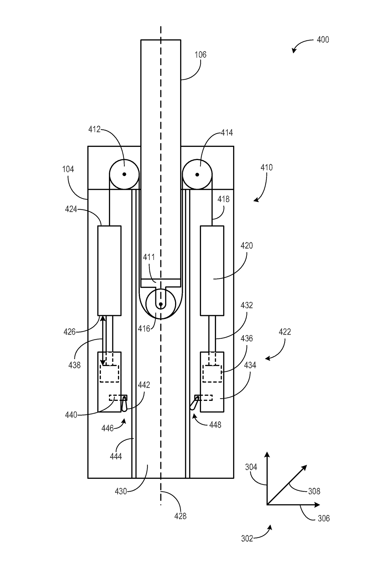
[0014]The following description relates to various embodiments of a weight adjustment system for a full-field breast ultrasound (FFBU) scanning apparatus. X-ray mammography is the most commonly used imaging method for mass breast cancer screening. However, x-ray mammograms only detect a summation of the x-ray opacity of individual slices over the entire breast. Alternatively, ultrasound imaging can separately detect sonographic properties of individual slices of breast tissue, thereby enabling users to detect breast lesions where x-ray mammography alone may fail.

[0015]Another well-known shortcoming of x-ray mammography practice is found in the case of dense-breasted women, including patients with high content of fibroglandular tissues in their breasts. Because fibroglandular tissues have higher x-ray absorption than the surrounding fatty tissues, portions of breasts with high fibroglandular tissue content are not well penetrated by x-rays and thus the resulting mammograms contain re...


PUM


Abstract

Various methods and systems are provided for imaging a tissue. In one example, a system comprises an adjustable arm, a scanning assembly attached to a first end of the adjustable arm, a counterweight coupled to a second end of the adjustable arm, and a weight adjustment system coupled to the counterweight and configured to modify an effective weight of the scanning assembly.

Description

FIELD[0001]Embodiments of the subject matter disclosed herein relate to medical imaging and the facilitation of ultrasonic tissue scanningBACKGROUND[0002]Volumetric ultrasound scanning of the breast may be used as a complementary modality for breast cancer screening. Volumetric ultrasound scanning usually involves the movement of an ultrasound transducer relative to a tissue sample and the processing of resultant ultrasound echoes to form a data volume representing at least one acoustic property of the tissue sample. Whereas a conventional two-dimensional x-ray mammogram only detects a summation of the x-ray opacity of individual slices of breast tissue over the entire breast, ultrasound can separately detect the sonographic properties of individual slices of breast tissue, and therefore may allow detection of breast lesions where x-ray mammography alone fails. Further, volumetric ultrasound offers advantages over x-ray mammography in patients with dense breast tissue (e.g., high co...

Claims


Application Information

Patent Timeline
Patent Type & Authority Patents(United States)
IPC IPC(8): A61B8/00A61B8/08
CPCA61B8/4218A61B8/0825A61B8/403A61B8/44
Inventor SUMMERS, DOUGLAS G.CHEN, JIAYU
Owner GENERAL ELECTRIC CO
Why Eureka
Try Eureka