Visualization of dual energy computed tomography airways data

a computed tomography and airway technology, applied in the field of visualizing airway trees, can solve the problems of scar tissue irritation and scar tissue not being distinguished from scar tissue using a standard ct image alone, and reducing the overall efficacy and convenience of using a dect-derived iodine map in a clinical and/or research environmen

Active Publication Date: 2017-03-28
SIEMENS HEALTHCARE GMBH
View PDF2 Cites 0 Cited by
  • Summary
  • Abstract
  • Description
  • Claims
  • Application Information

AI Technical Summary

Benefits of technology

[0007]In accordance with the present teachings, a visualization of a patient's airway walls from a computed iodine map obtained by dual energy computed tomography (DECT) is provided. The visualization derived from the computed iodine map may be presented in a bronchogram-like format familiar to radiologists. The bronchogram-like format is referred to herein as a bronchodine (e.g., a composite word derived from “bronchogram” and “iodine”), and depicts iodine concentration in regions of diagnostic interest (e.g., airway walls, etc.) while excluding extraneous regions (e.g., connective tissue, etc.) that are deemed distracting and unnecessary to diagnosis and / or evaluation. Methods in accordance with the present teachings may visualize an entire DECT dataset in 3D with minimal post-processing, thereby retaining a substantial portion of the original data.

Problems solved by technology

A thickening in the walls of the airways may be a result of scar tissue (e.g., permanent damage) or a potentially treatable tissue irritation.
However, it may not be possible to distinguish scar tissue from tissue irritation using a standard CT image alone.
This extraneous information may reduce the overall efficacy and convenience of using a DECT-derived iodine map in a clinical and / or research environment.
Although sophisticated post-processing methods may be useful for simplifying or correcting views, computation errors may be introduced as a result, thereby compromising the interpretation of the data.
Additionally, the illustrations are merely representational and may not be drawn to scale.

Method used


Image

  • Visualization of dual energy computed tomography airways data
  • Visualization of dual energy computed tomography airways data
  • Visualization of dual energy computed tomography airways data

Examples


Embodiment Construction

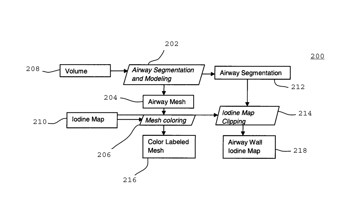
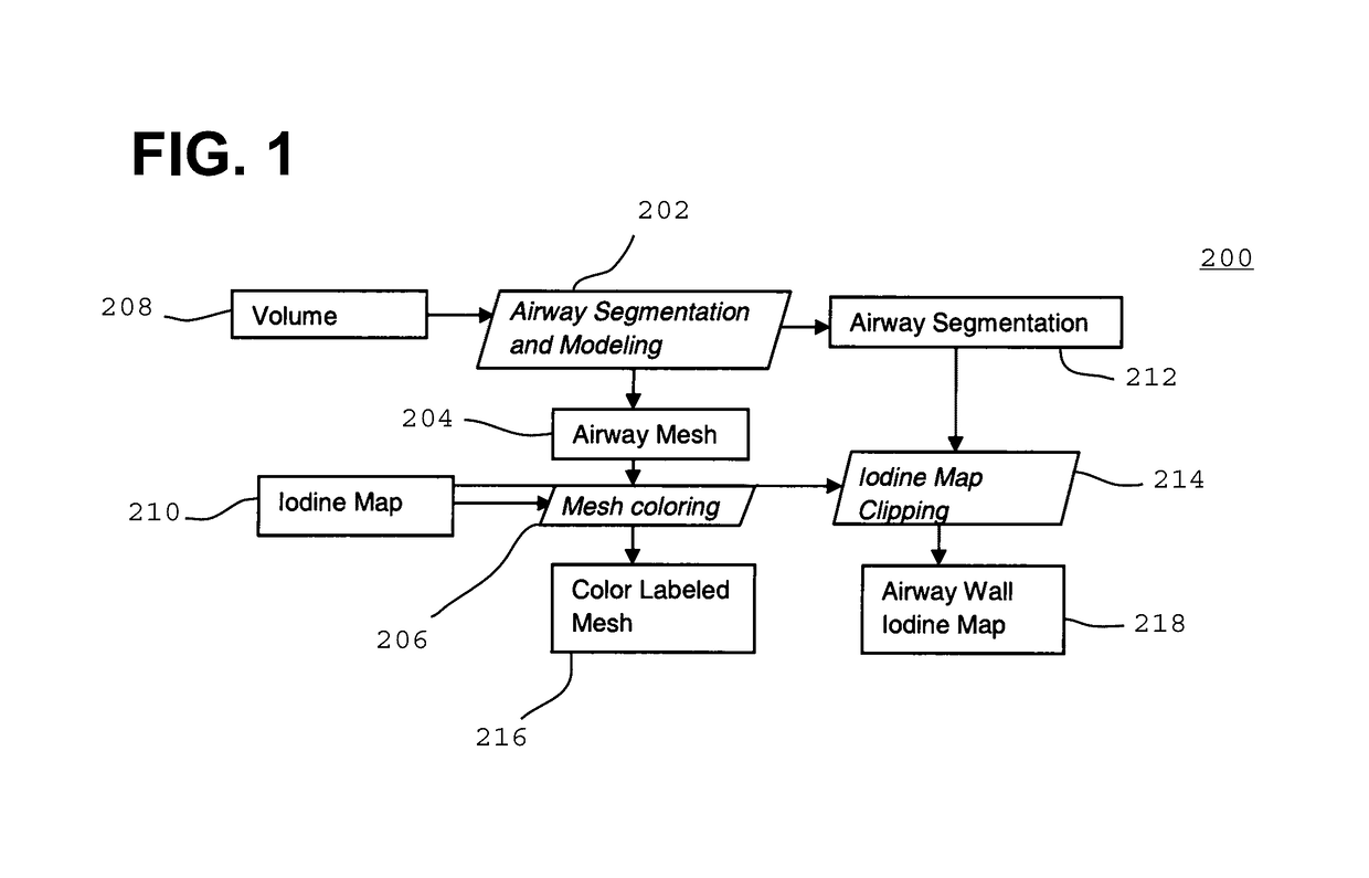
[0018]To improve the phenotyping of airway diseases, blood perfusion may be measured via concentration of a contrast agent (e.g., iodine) using DECT imaging. The iodine maps thus obtained may be used to differentiate between scar tissue and inflamed airway walls. This level of differentiation may be helpful in indicating an optimum course of treatment for various pulmonary diseases, including but not limited to bronchiectasis, asthma, cystic fibrosis, and chronic obstructive pulmonary disease (COPD).

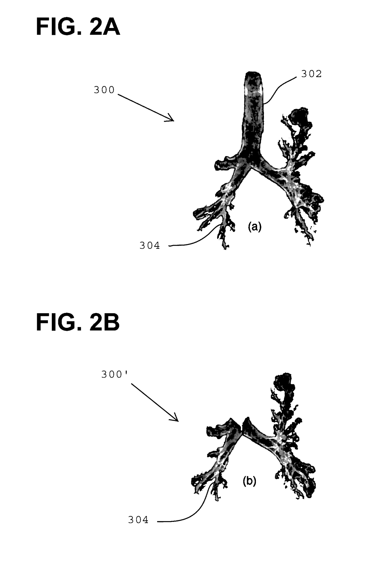
[0019]Iodine maps derived from DECT data may be useful in assessing airway morphology and blood perfusion in patients. In accordance with the present teachings, techniques for visualizing iodine maps that selectively focus on the airways of the patient have been developed and are described herein. In some embodiments, the methods may provide a bronchogram-like view of a patient's airway tree in a format that is familiar to radiologists. A conventional bronchogram is a radiograph of the b...


PUM


Abstract

A method for depicting an airway tree of a patient includes: (a) generating an iodine map of the airway tree from dual energy computed tomography (DECT) imaging data acquired from the patient; (b) defining a region of interest of the airway tree from the DECT imaging data; (c) rendering at least a portion of the airway tree based on information derived from the iodine map and the defined region of interest; and (d) displaying a graphical image of at least a portion of the airway tree on a user interface. Systems for depicting an airway tree of a patient are described.

Description

RELATED APPLICATIONS[0001]This claims the benefit of U.S. Provisional Application No. 61 / 882,132, filed Sep. 25, 2013. The entire contents of the provisional application are incorporated herein by reference, except that in the event of any inconsistent disclosure or definition from the present specification, the disclosure or definition herein shall be deemed to prevail.TECHNICAL FIELD[0002]The present teachings relate generally to the visualization of airway trees and, in some embodiments, to the diagnosis and / or evaluation of pulmonary disease using visualizations of a patient's airway tree.BACKGROUND[0003]A thickening in the walls of the airways may be a result of scar tissue (e.g., permanent damage) or a potentially treatable tissue irritation. The airways in the lungs of a patient may be examined for anatomical defects using standard computed tomography (CT). However, it may not be possible to distinguish scar tissue from tissue irritation using a standard CT image alone.[0004]...

Claims


Application Information

Patent Timeline
Patent Type & Authority Patents(United States)
IPC IPC(8): A61B6/00A61B6/03G06T7/00
CPCA61B6/466A61B6/032A61B6/4241A61B6/469A61B6/482A61B6/50G06T7/136G06T2207/10081G06T2207/10152G06T2207/30061G06T7/11
Inventor KIRALY, ATILLA PETERODRY, BENJAMIN L.NOVAK, CAROL L.
Owner SIEMENS HEALTHCARE GMBH
Why Eureka
Try Eureka