
Method of detecting movement between an implant and a bone

a technology of bone and implant, applied in the field of bone implants, can solve the problems of time-consuming process and the general inability to provide the desired accuracy of surgical implantation of markers

Active Publication Date: 2017-04-18
HALIFAX BIOMEDICAL
View PDF6 Cites 19 Cited by
  • Summary
  • Abstract
  • Description
  • Claims
  • Application Information

AI Technical Summary

Problems solved by technology

Markers are currently implanted during surgery; however, this process can be time consuming.
In addition, the surgical implantation of markers generally does not necessarily provide the desired accuracy.

Method used


Image

  • Method of detecting movement between an implant and a bone
  • Method of detecting movement between an implant and a bone
  • Method of detecting movement between an implant and a bone

Examples


Embodiment Construction

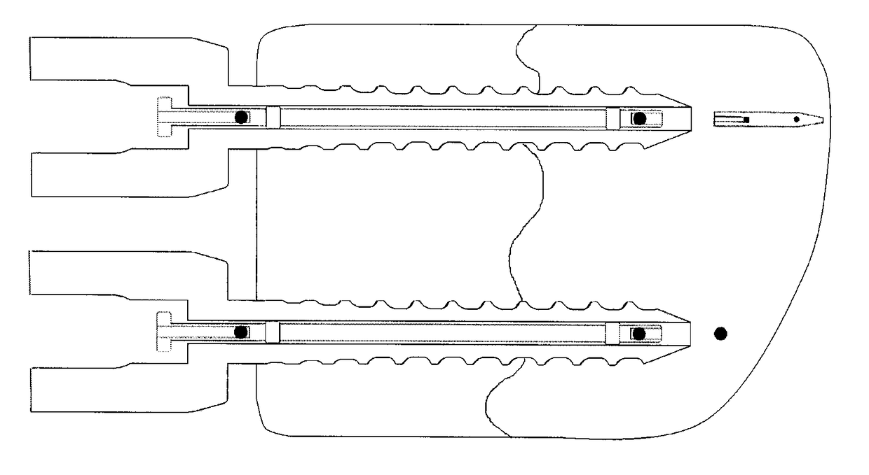
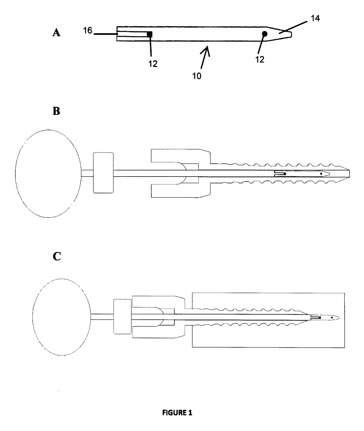
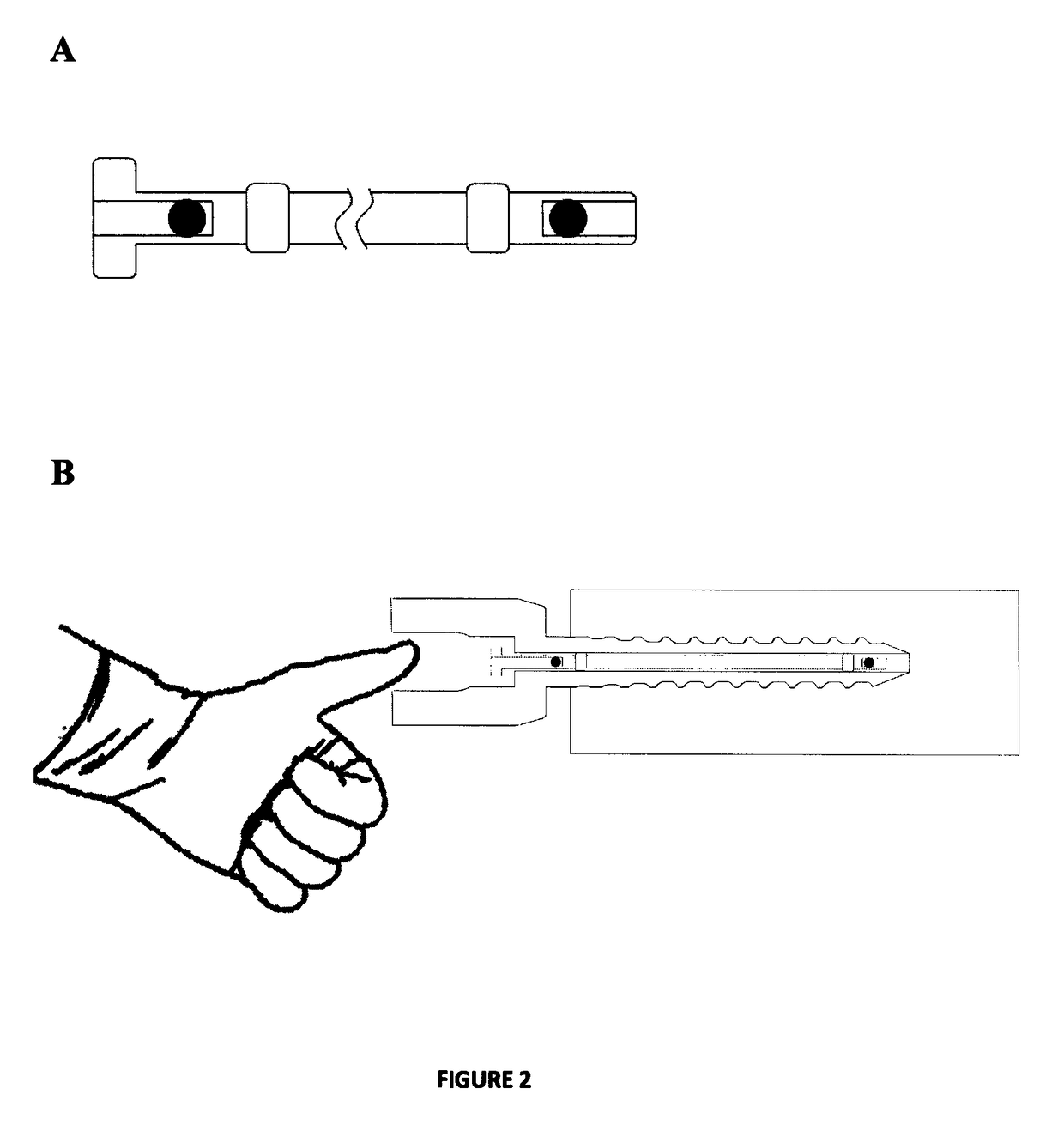
[0022]A method to detect movement between a cannulated bone implant and a bone in a patient is provided by measuring the relative position of the bone, in which one or more radiopaque bone markers have been implanted through the implant within the bone and one or more radiopaque implant markers have been implanted into the implant. The method comprises determining a first relative position of the bone marker to the implant marker under a first parameter; and determining a second relative position of the bone marker to the implant marker under a second parameter, wherein a change in the position of the bone marker relative to the implant marker from the first relative position to the second relative position is indicative of movement of either the implant or the bone.

[0023]In order to conduct the present method, one or more radiopaque markers are implanted into the bone through a cannulated implant within the bone. The use of a cannulated implant provides a means of delivering one or...


PUM


Abstract

A method of measuring the relative position of a bone implant within a bone in a patient to detect movement between the implant and bone is provided, wherein one or more radiopaque bone markers have been implanted into the bone through a cannulated implant within the bone and one or more radiopaque implant markers have been implanted into the implant. The method comprises: i) determining a first relative position of the bone marker to the implant marker under a first parameter; and ii) determining a second relative position of the bone marker to the implant marker under a second parameter, wherein a change in the position of the bone marker relative to the implant marker from the first relative position to the second relative position is indicative of movement of either the implant or the bone.

Description

[0001]This application is a 371 national stage application of PCT Patent Application No. PCT / CA2011 / 000967, filed Aug. 25, 2011, entitled “A Method of Detecting Movement Between an Implant and a Bone,” which claims priority to U.S. Provisional Application No. 61 / 376,916, filed Aug. 25, 2010, the content of all of which is hereby incorporated in its entirety by reference.FIELD OF THE INVENTION[0002]The present invention relates generally to the field of orthopedics, and in particular, to bone implants and the use of bone implants in imaging methods.BACKGROUND[0003]In orthopedics there is value in determining the relative motion of two or more rigid bodies such as bone segments and bone implants. X-ray imaging is generally used to determine relative position and orientation of bone structures and implants. Radiostereometric analysis or roentgen stereophotogrammetric analysis (RSA) is a method of obtaining two simultaneous x-ray exposures of the same implant / bone structure to determine...

Claims


Application Information

Patent Timeline
Patent Type & Authority Patents(United States)
IPC IPC(8): A61B5/103A61B6/02A61B6/00A61F2/46A61B17/86A61B17/70A61B6/12A61B90/00
CPCA61B6/022A61B6/486A61B6/505A61B6/5229A61B90/39A61B6/12A61B17/7032A61B17/864A61B2090/061A61B2090/0812A61B2090/3904A61B2090/3916A61B2090/3966A61B2090/3987A61F2002/4658
Inventor MUNRO, CHAD R.VAN DER PUT, RICHARD W.BURGER, ANDREAS
Owner HALIFAX BIOMEDICAL
Learn More