Sealing device for sealing a passage for a medical instrument

a sealing device and medical instrument technology, applied in the field of sealing devices for sealing passages of medical instruments, can solve problems such as similar problems, and achieve the effects of reducing the risk of damage to the ring seal or the slit membrane, low force, and high degree of reliability

Active Publication Date: 2017-04-18
KARL STORZ GMBH & CO KG
View PDF32 Cites 8 Cited by
  • Summary
  • Abstract
  • Description
  • Claims
  • Application Information

AI Technical Summary

Benefits of technology

[0027]The increased wall thickness in the transition area of the sealing device results in increased stiffness of the transition area. This increased stiffness in the transition area reduces deformations of the sealing device in the transition area. An increased stiffness of the transition area reduces the risk of the sealing device turning inside out starting from the transition area. A medical instrument in the sealing device can therefore be moved in both directions, i.e. both in the distal direction and also in the proximal direction, with less risk of the sealing device turning inside out.
[0053]In particular, a friction-reducing layer is provided on the entire surface of the sealing device or at least in the area of the ring seal and / or of the slit membrane. A friction-reducing layer can reduce the forces that act when inserting an instrument into the sealing device and when withdrawing the instrument from the sealing device. The layer can reduce both the static friction and also the kinetic friction. A reduction of the kinetic and / or static friction can allow a more sensitive movement of a medical instrument in the sealing device and can reduce mechanical loading of the sealing device. Reduced mechanical loading of the sealing device can reduce the risk of the first wall turning inside out and the risk of the sealing device suffering damage, for example tearing.

Problems solved by technology

Partially similar problems may arise in a work channel of an endoscope or when inserting a catheter into a blood vessel.

Method used


Image

  • Sealing device for sealing a passage for a medical instrument
  • Sealing device for sealing a passage for a medical instrument
  • Sealing device for sealing a passage for a medical instrument

Examples


Embodiment Construction

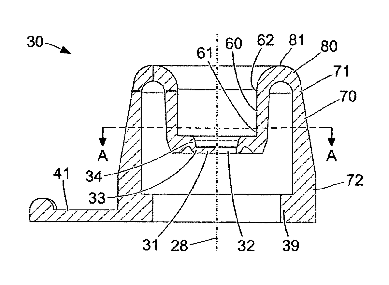
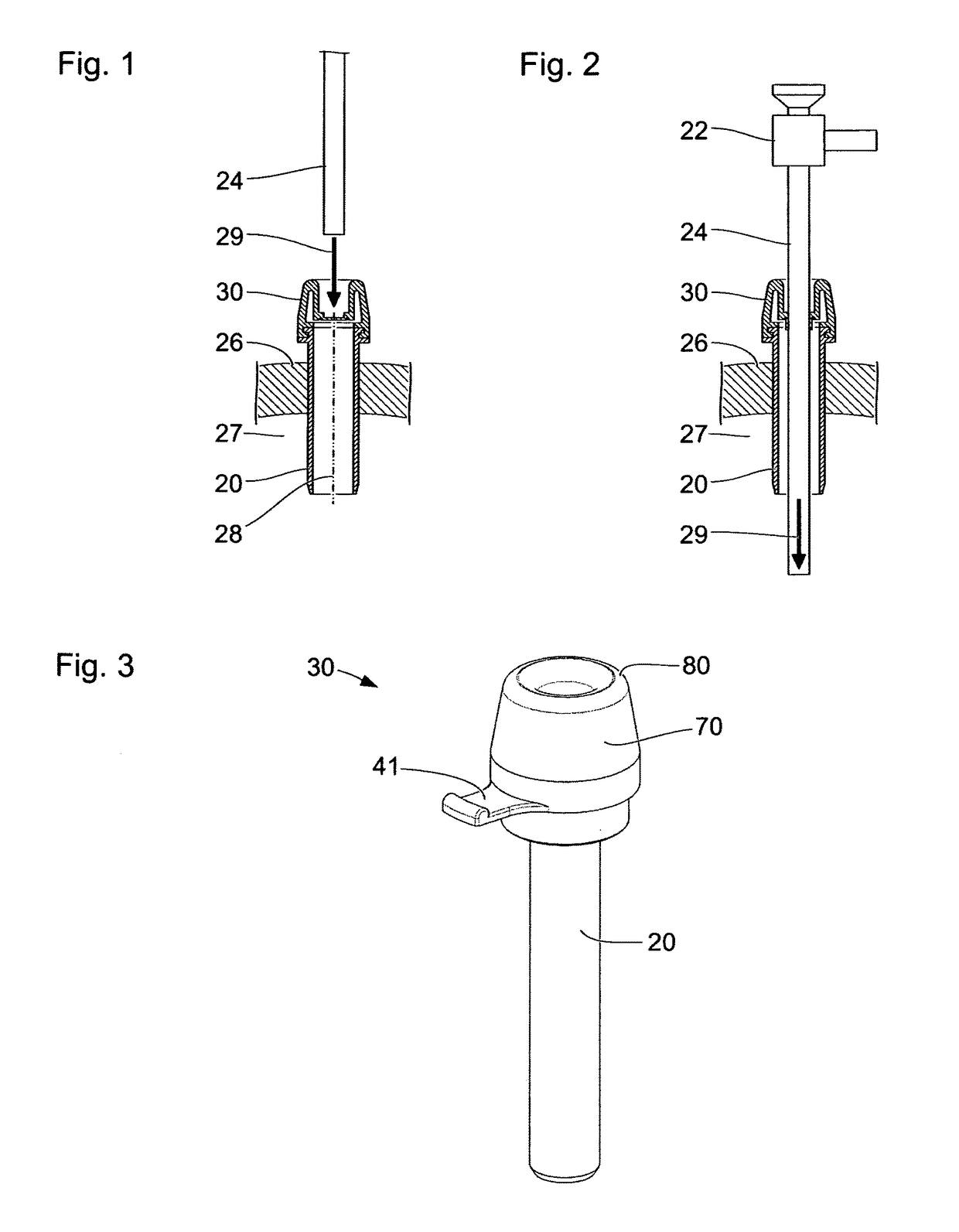
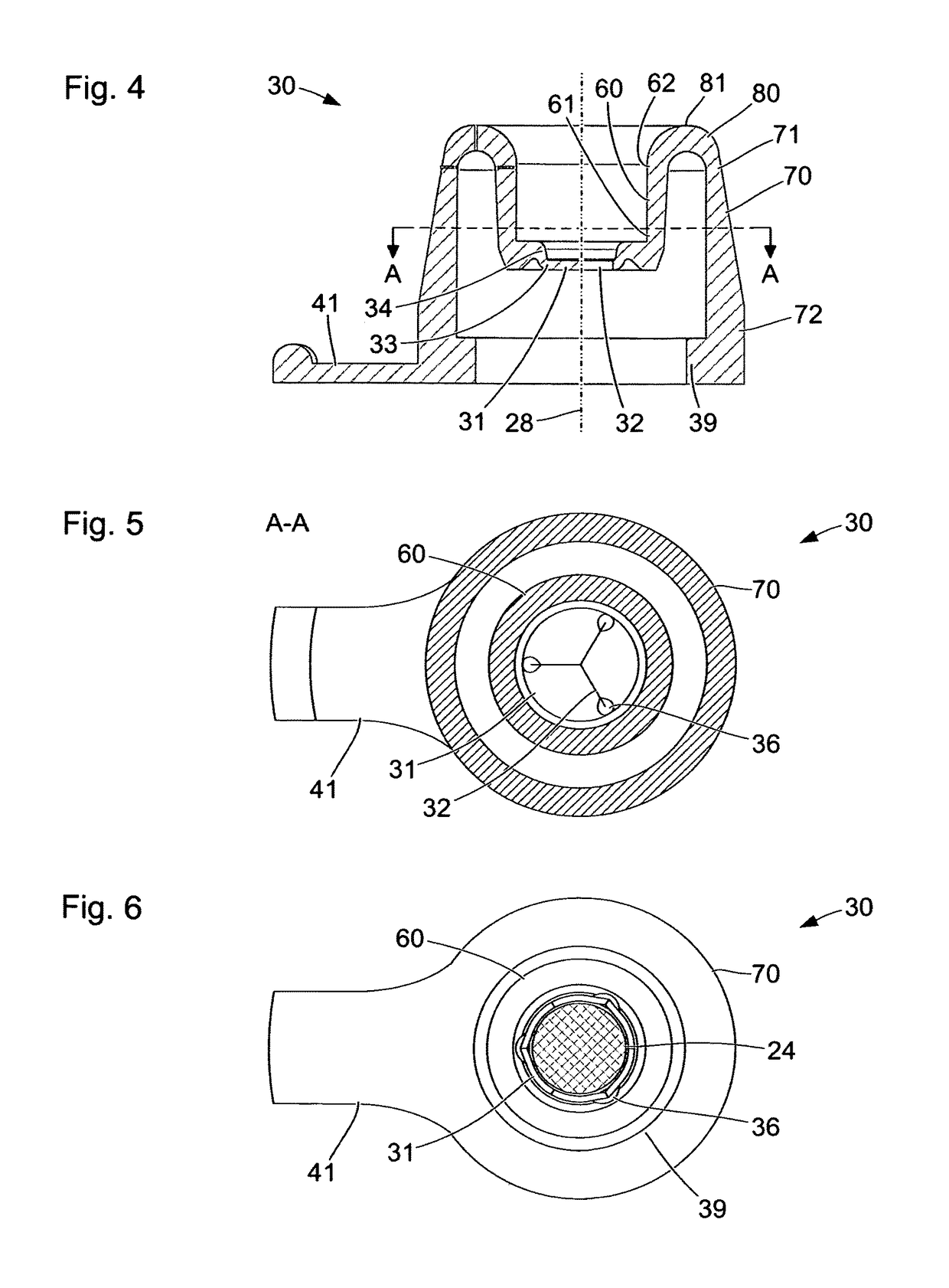
[0068]FIGS. 1 and 2 show schematic sectional views of a tube 20 of a trocar, which is fitted into an abdominal wall 26 of a patient. Through the lumen of the tube 20, one or more medical instruments can be inserted into a cavity 27 under the abdominal wall 26 of the patient. The tube 20, the abdominal wall 26 and the cavity 27 under the abdominal wall 26 are shown in a section along a plane containing a longitudinal axis 28 of the tube 20. In particular, the tube 20 is at least in part rotationally symmetrical with respect to the longitudinal axis 28. The tube 20 comprises a sealing device 30 at its proximal end outside the cavity 27. Embodiments of the sealing device 30 are described in detail with reference to FIGS. 3 to 8.

[0069]FIGS. 1 and 2 also show a medical instrument 22 with a shank 24. Since the internal design of the medical instrument 22 and of the shank 24 thereof are not relevant as regards the properties of the tube 20 that are described below, only contours of the med...


PUM


Abstract

A sealing device for a medical instrument includes a fastening area and a sealing area designed to either bear on the circumference of a medical instrument inserted into the sealing device or to close the sealing device when no medical instrument is inserted. The sealing device further includes a first elastic wall enclosing the sealing area in a ring shape, with an annular first edge, which is connected to the sealing area, and an annular second edge, and a second elastic wall which encloses the first wall in a ring shape and is spaced apart there from, with an annular first edge and an annular second edge, which is connected to the fastening area.

Description

FIELD OF THE INVENTION[0001]The present invention relates to a sealing device for sealing a passage for a medical instrument, in particular for sealing a tube through which an endoscope and / or other medical instruments can be inserted into a natural or artificial body cavity in a microinvasive medical intervention, or for sealing a work channel of an endoscope.BACKGROUND OF THE INVENTION[0002]Laparoscopy is one example of a microinvasive medical method. By means of a trocar, an artificial access route to the abdominal cavity of a patient is created through the abdominal wall. Through the lumen of a tube of the trocar that remains in the abdominal wall during the laparoscopic intervention, it is possible for an endoscope and / or other medical instruments (for example forceps, scissors, needle holders) to be inserted into the abdominal cavity. During the laparoscopy, the abdominal space is filled carbon dioxide or another gas in order to create a pneumoperitoneum, a hollow space permit...

Claims


Application Information

Patent Timeline
Patent Type & Authority Patents(United States)
IPC IPC(8): A61B17/34A61B17/00
CPCA61B17/00234A61B17/3462A61B17/3423A61B2017/3419A61B2017/3464
Inventor OBERLAENDER, MARTINWAGNER, SEBASTIAN
Owner KARL STORZ GMBH & CO KG
Why Eureka
Try Eureka