Methods and compositions for somatic cell proliferation and viability

a somatic cell and composition technology, applied in the field of somatic cell proliferation and viability, can solve the problems of stem cell and progenitor cell loss, stem cell function and proliferative capacity of adult stem cells, and limited regeneration capacity of somatic cells, so as to increase and improve the viability of the neuron

Active Publication Date: 2017-09-12
RGT UNIV OF CALIFORNIA
View PDF5 Cites 1 Cited by
  • Summary
  • Abstract
  • Description
  • Claims
  • Application Information

AI Technical Summary

Benefits of technology

[0005]Muscle tissue in adult vertebrates regenerates from reserve myoblasts called satellite cells. Satellite cells are distributed throughout muscle tissue and are mitotically quiescent in the absence of injury or disease. Following recovery from damage due to injury or disease or in response to stimuli for growth or hypertrophy, satellite cells reenter the cell cycle, proliferate and enter existing muscle fibers or undergo differentiation into multinucleated myotubes, which form new muscle fiber. The myoblasts ultimately yield replacement muscle fibers or fuse into existing muscle fibers, thereby increasing fiber girth.
[0021]Methods are also provided for screening of other agents that can improve somatic cell proliferation and / or increase viability of somatic cells in the presence of heparin binding protein composition.

Problems solved by technology

Unlike embryonic stem cells, somatic cells have a limited capacity for regeneration.
Although, stem cells and progenitor cells present in organs of an adult organism can proliferate and lead to generation of new somatic cells to replace older, injured, diseased, dying, or dead somatic cells, the stem cells and progenitor cells may lose this capacity due to a number of causes.
With explantation and in vitro culture, adult stem and progenitor cells lose stem cell function and proliferative capacity, and suffer high mortality when reintroduced in vivo.

Method used


Image

  • Methods and compositions for somatic cell proliferation and viability
  • Methods and compositions for somatic cell proliferation and viability
  • Methods and compositions for somatic cell proliferation and viability

Examples


example 1

mTeSR-1 Growth Medium has Pro-myogenic Activity, which is Due to the High Levels of FGF-2, and hESC-secreted Factors Act Independently of Recombinant FGF-2

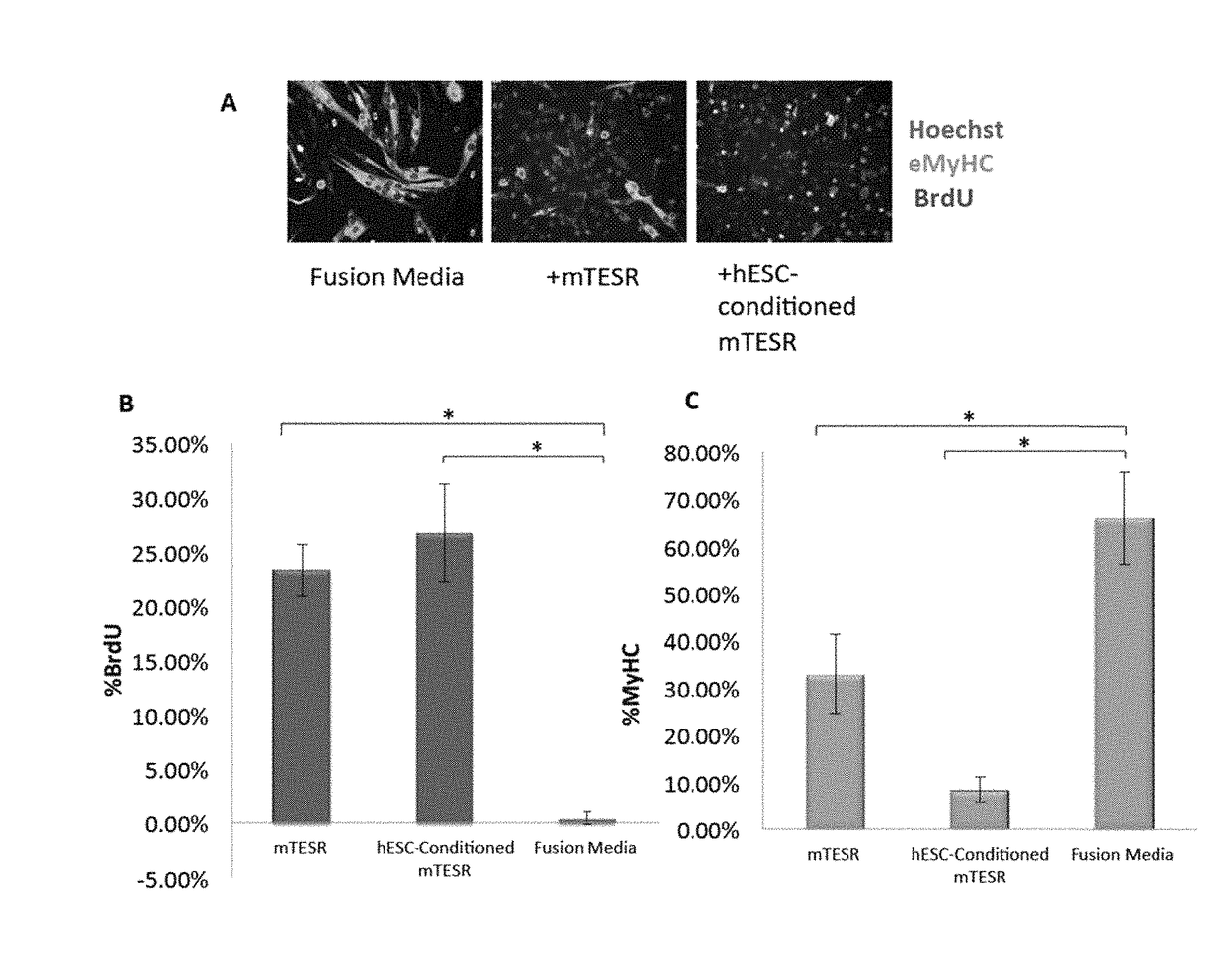
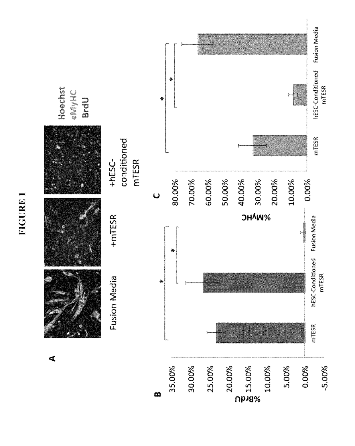
[0148]Our previous work established that injection of hESCs—which were cultured on mouse embryonic fibroblasts (MEF) and in standard, highly mitogenic, embryonic cell growth medium—enhanced old muscle regeneration (Carlson, M. E. and I. M. Conboy, Aging Cell, 2007. 6(3): p. 371-82). In our more recent work, the hESCs have been cultured in mTeSR-1 (Stem Cell Technologies), a defined feeder-free medium which is also highly mitogenic (Ludwig, T. E., et al., Nat Methods, 2006. 3(8): p. 637-646), and we investigated whether and to what degree the pro-myogenic effects of hESC-conditioned medium was due to the residual activity of the hESC growth / expansion medium. Primary muscle progenitor cells (myoblasts) were cultured overnight in a mitogen-low fusion medium that typically induces differentiation of myoblasts into multinucleated eMyHC...

example 2

FGF-2 Signaling and Satellite Cell Proliferation are not Increased with Age

[0150]FGF-2, which often functions as a mitogen, was recently reported to contribute to the aging and depletion of mouse satellite cells. However, the canonical model of muscle stem cell aging postulates that a decline in such mitogens over time leads to reduced activation of satellite cells that are resident to old tissue (Conboy, I. M. and T. A. Rando, Cell Cycle, 2012. 11(12): p. 2260-7; Grounds, M.D., Ann. N.Y. Acad. Sci., 1998. 854: p. 78-91; onboy, I. M. and T. A. Rando, Cell Cycle, 2005. 4(3): p. 407-410), so we explored these phenomena in more detail. The levels of FGF-2 were determined by Western Blotting in muscle fibers that were derived from Tibialis Anterior (TA) and Gastrocnemius (Gastroc) muscle of young and old mice. As shown in FIG. 3A (quantified in 3B), a significant increase in FGF-2 protein was observed with age in myofibers, consistent with Chakkalakal et al. FGF-2 signals through the MA...

example 3

The Pro-regenerative Activity of hESC-Secreted Factors is Contained in Proteins with Heparin Binding Domains

[0153]To confirm that the factors in hESC conditioned medium were proteins, hESC conditioned Opti-MEM was treated with proteinase-K agarose beads, and the beads were removed before mixing 50 / 50 with Opti-MEM and 5% mouse serum, for culture with injury-activated satellite cells with associated fibers from old muscle, as above. All proliferative activity of the conditioned medium was lost after proteinase treatment, indicating that protein(s) conferred the pro-regenerative activity (FIG. 7).

[0154]To deplete heparin-binding proteins, hESC-conditioned medium was incubated with heparin binding domain-coated acrylic beads. Muscle progenitor cells were then cultured in this heparin-depleted hESC-conditioned medium, hESC-conditioned medium, or controls (medium alone and medium conditioned by differentiated cells that lack the pro-regenerative activity). Proliferation of primary muscle...


PUM

PropertyMeasurementUnit
timeaaaaaaaaaa
timeaaaaaaaaaa
timeaaaaaaaaaa

Abstract

Methods and compositions for somatic cell proliferation as well as increasing viability of somatic cells are provided. The compositions include heparin binding protein isolated from a medium conditioned by growth of pluripotent stem cells, such as, human embryonic stem cells, human embryonic carcinoma cells. The methods include contacting a somatic cell with a heparin binding protein composition for a sufficient period of time to provide for enhanced proliferation and / or viability of the somatic cell as compared to the absence of the heparin binding protein composition.

Description

CROSS-REFERENCE TO RELATED APPLICATION[0001]Pursuant to 35 U.S.C. §119(e), this application claims priority to the filing date of U.S. Provisional Application No. 61 / 828,543, filed May 29, 2013, the disclosure of which is incorporated herein by reference.STATEMENT REGARDING FEDERALLY SPONSORED RESEARCH[0002]This invention was made with government support under Grant No. AG027252 awarded by National Institutes of Health. The government has certain rights in the invention.INTRODUCTION[0003]Unlike embryonic stem cells, somatic cells have a limited capacity for regeneration. While, embryonic stem cells have generated considerable interest, the stem cells resident in differentiated tissues may also provide an important source of regenerative capacity. These somatic, or adult, stem cells are undifferentiated cells that reside in differentiated tissues, and have the properties of self-renewal and generation of differentiated cell types. The differentiated cell types may include all or some...

Claims


Application Information

Patent Timeline
Patent Type & Authority Patents(United States)
IPC IPC(8): C12N5/00C12N5/077C12N5/0793
CPCC12N5/0619C12N5/0659C12N2501/91C12N2502/02
Inventor CONBOY, IRINA M.CONBOY, MICHAEL J.YOUSEF, HANADIESCHAFFER, DAVID V.
Owner RGT UNIV OF CALIFORNIA
Why Eureka
Try Eureka