
Image monitoring device and method for damage on skin and subcutaneous tissue

A technology for image monitoring and subcutaneous tissue, applied in the directions of ultrasound/acoustic/infrasonic image/data processing, organ movement/change detection, diagnosis, etc., can solve the problem that there is no effective monitoring of skin and subcutaneous tissue damage

Active Publication Date: 2008-11-05
RONGHAI SUPERSONIC MEDICINE EN
View PDF4 Cites 1 Cited by
  • Summary
  • Abstract
  • Description
  • Claims
  • Application Information

AI Technical Summary

Problems solved by technology

[0005] There is no effective way to monitor skin and subcutaneous tissue damage during current HIFU therapy

Method used


Image

  • Image monitoring device and method for damage on skin and subcutaneous tissue
  • Image monitoring device and method for damage on skin and subcutaneous tissue

Examples


Embodiment Construction

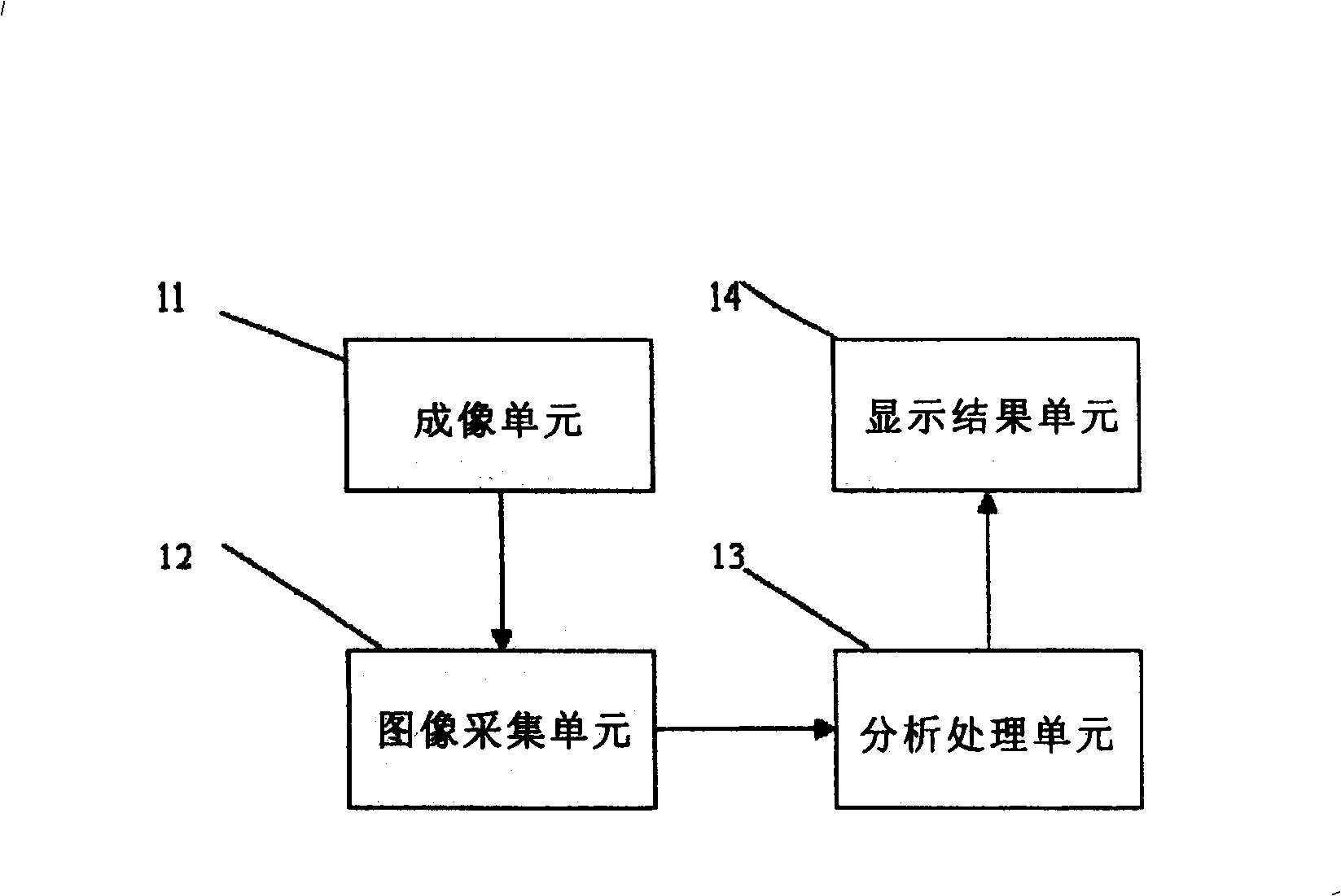
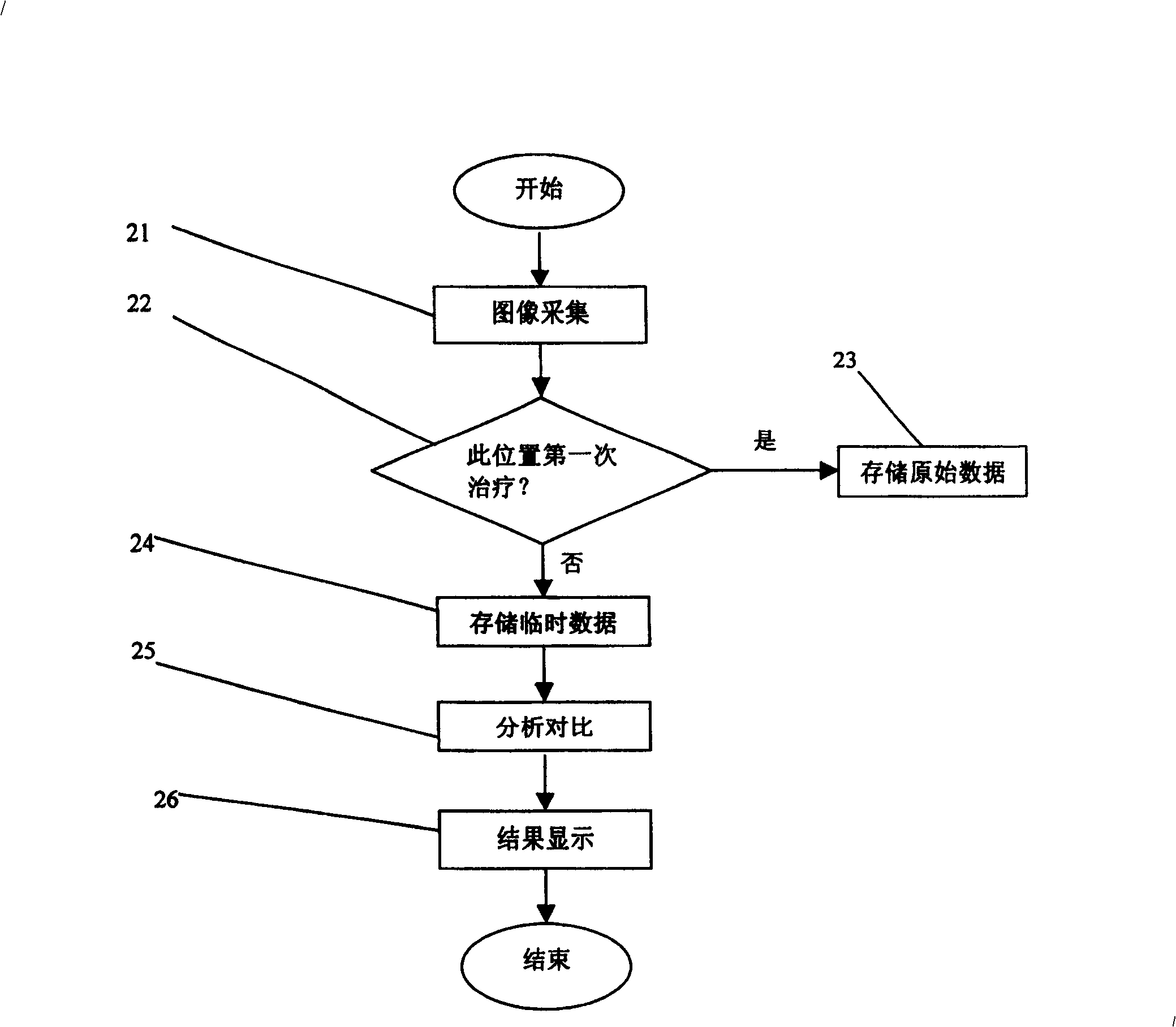
[0011] Use the following figure 1 and figure 2 , the device and method for image monitoring of skin and subcutaneous tissue damage of the present invention will be described. figure 1 is a structural block diagram of the skin and subcutaneous tissue damage image monitoring device of the present invention, figure 2 It is a flowchart of image monitoring of skin and subcutaneous tissue damage in the present invention.

[0012] Such as figure 1 As shown, the image monitoring device for skin and subcutaneous tissue damage of the present invention includes an imaging unit 11 , an image acquisition unit 12 , an analysis and processing unit 13 and a result display unit 14 . Wherein, the imaging unit 11 is a device for obtaining images of the skin and subcutaneous tissue within the ultrasound channel range, such as B-ultrasound, CT or MRI (magnetic resonance imaging) and other devices. The image acquisition unit 12 is connected with the imaging unit 11, and is used to acquire ima...


PUM


Abstract

An image monitor used in high-intensity focused ultrasonic therapy for real-time monitor to the injury of skin and hypoderm is composed of an imaging unit for taking the images of skin and hypoderm, an image acquiring unit for acquiring the data about said images, an analyzing and processing unit for storing said image data, comparing the data before treating with the data uring treating, and analyzing the injury, and a result display unit. Its method is also disclosed.

Description

field of invention [0001] The invention relates to an image monitoring device for skin and subcutaneous tissue damage, in particular to an image monitoring device for skin and subcutaneous tissue damage during high-intensity focused ultrasound treatment. Background technique [0002] Medical research has found that the heat resistance of tumor cells is worse than that of normal cells. In a temperature environment above 42.5°C, tumor cells die within 30 minutes, while normal cells are slightly damaged and reversible. Based on this characteristic of tumor cells, the high-intensity focused ultrasound tumor therapy system uses ultrasound as the energy source, uses its penetrability and focusability, and couples the ultrasound with low average sound intensity emitted by the probe through the medium and passes through the skin. Enter the human tumor tissue, focus or converge to a spatial point to form an average sound intensity of 1000w / m 2 The above focal region (focal region si...

Claims


Application Information

Patent Timeline
Patent Type & Authority Patents(China)
IPC IPC(8): A61B8/00A61B8/08A61B8/13
Inventor 陈文直木木王明胜文银刚汪海
Owner RONGHAI SUPERSONIC MEDICINE EN
Learn More