Preparation method of glue adhesion amnion

A technology of gluing amniotic membrane and amniotic membrane, applied in medical science, prosthesis, etc., can solve the problems that it is difficult to fully exert various biological activities, sutures are easy to induce inflammation, and amniotic membrane dissolves and falls off, so as to inhibit new blood vessels and scars The effect of growing, overcoming easy tearing, and easy cutting and suturing

Inactive Publication Date: 2009-04-22
CHONGQING MEDICAL UNIVERSITY
View PDF4 Cites 2 Cited by
  • Summary
  • Abstract
  • Description
  • Claims
  • Application Information

AI Technical Summary

Problems solved by technology

[0004] At present, most of the amniotic membranes used clinically are single-layer amniotic membranes, but the single-layer amniotic membranes have one of the biggest disadvantages: after amniotic membrane transplantation, the amniotic membranes generally can only be kept on the wound for a few days to two weeks, and finally completely dissolve and fall off. It is difficult to fully exert its various biological activities, so it cannot produce long-lasting biological therapeutic effects
But all the methods are to overlap the amniotic membrane, suture and fix it on the ocular surface or stuff it on the wound surface by using the suture method. After this method, the patient needs to pressurize for a long time or wear a hydrophilic corneal contact. However, even so, the existing double or multi-layer amniotic membranes are easily delaminated, forming interlaminar effusion, and the superficial amniotic membranes fall off quickly, and then all the amniotic membranes dissolve and fall off, and the overlapping of the amniotic membranes increases the difficulty of the operation, requiring a large number of sutures. New damage is added, and too many sutures can easily induce inflammation and neovascularization

Method used

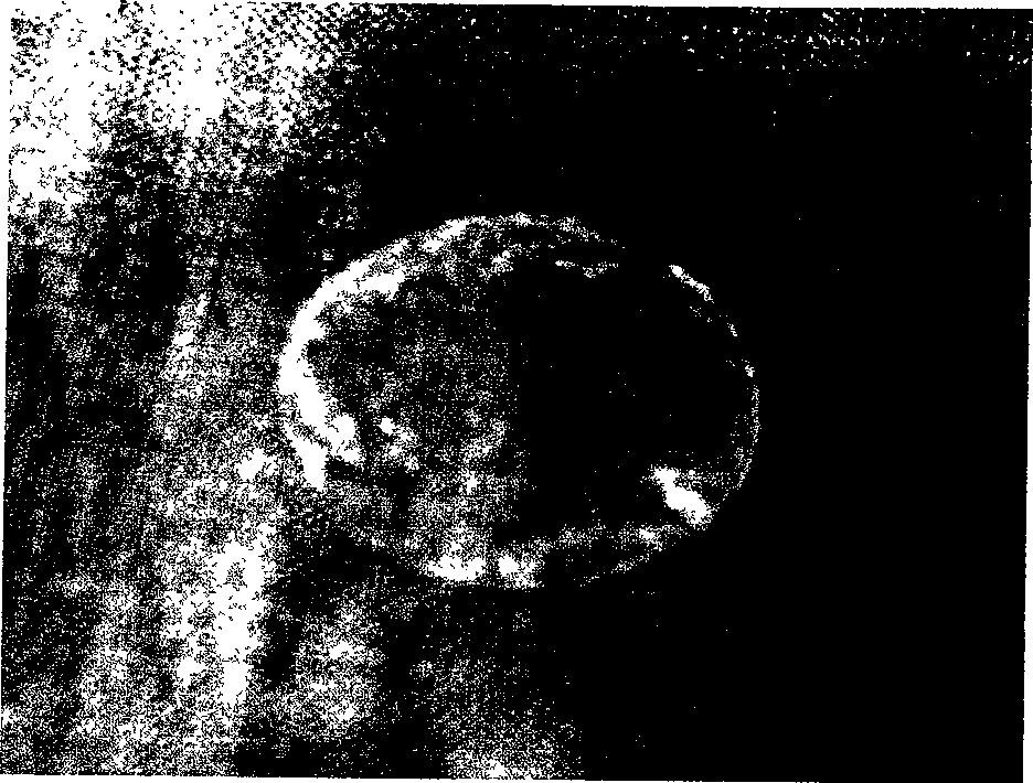
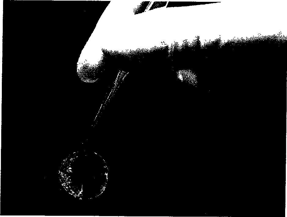

Image

  • Preparation method of glue adhesion amnion
  • Preparation method of glue adhesion amnion
  • Preparation method of glue adhesion amnion

Examples


Embodiment 1

[0024] Embodiment 1 Preparation of double-layer glued amniotic membrane

[0025] 1) Take a healthy cesarean section placenta, wash it with medical saline containing 2000U / ML gentamicin and 2.5UG / ML amphotericin B, and bluntly separate it through the potential gap between the amnion and chorion to obtain a smooth and transparent , Avascular amniotic membrane, and sterilized by immersing in anti-biological saline mixture for 20 minutes.

[0026] 2) Preparation of de-epithelialized amniotic membrane: spread the epithelial surface of the amniotic membrane upward on nitrocellulose filter paper (0.45um micropore); digest the amniotic membrane with 0.25% trypsin and 0.06% EDTA in a 37°C incubator for 30 minutes, and then use The residual epithelium was scraped off with a cell brush, and a small amount of de-epithelized amniotic membrane was randomly taken, and the epithelial cells were scraped clean under a light microscope.

[0027] 3) Double-layer amniotic membrane glue connection...

Embodiment 2

[0030] Embodiment 2 Preparation of double-layer gel-linked amniotic membrane

[0031] The acquisition and processing method of the amnion is the same as the step 1) of embodiment 1; after the fresh amnion epithelial surface that has been processed is pasted on the nitrocellulose filter paper smoothly, then the fibrin glue is evenly sprayed on the basement membrane surface of the amnion, and Paste the other amniotic membrane flatly on it. At this time, the nitrocellulose filter paper sandwiches the two layers of amniotic membrane in the middle, and gently press it with a flat-bottomed vessel for 3 minutes to make the amniotic membrane adhere tightly. The surface of the basement membrane is attached to the surface of the basement membrane).

[0032] The glue-linked amniotic membrane prepared according to the above-mentioned embodiment 1 and implementation 2 is double-layer fresh glue-linked amniotic membrane, which can be directly applied in clinic. If you want to store it, put...

Embodiment 3

[0033] Example 3 Preparation of Double-Layer Adhesive-Linked Amnion Using Glycerol-Preserved Monolayer Amnion

[0034] The acquisition and processing method of the amniotic membrane is the same as step 1) of Example 1. The treated fresh single-layer amniotic membrane is soaked and dehydrated in pure glycerin for 24 hours, and then transferred to another sterile vial filled with pure glycerin and sealed at 4°C. stand-by. When preparing glue-linked amniotic membrane, take out the single-layer amniotic membrane preserved in glycerin, rehydrate with sterile saline or equilibrium solution for 30 minutes, and fully wash off the glycerol on the amniotic membrane surface, according to step 2) in Example 1. Methods The epithelial amniotic membrane was prepared. Concrete gluing mode is described in step 3) or embodiment 2 with embodiment 1. The prepared gel-linked amniotic membrane can be stored continuously, and after being dehydrated with pure glycerin for 24 hours, it is then trans...


PUM

PropertyMeasurementUnit
thicknessaaaaaaaaaa

Abstract

The invention relates to an adhesive amniotic membrane, which is a double layer or multi-layer adhesive amniotic membrane bonded with amniotic membrane and fibrin albumen glue. The preparing method comprises: bonding the fresh amniotic membrane of epithelium removed with fresh amniotic membrane without removing epithelium by using fibrin albumen glue; or condensing the amniotic membrane immersing in the glycerol, and then bonding the fresh amniotic membrane of epithelium removed with fresh amniotic membrane without removing epithelium by using fibrin albumen glue; or bonding with the fresh amniotic membrane without removing epithelium by using fibrin albumen glue; finally putting the bonded adhesive amniotic membrane into pure glycerol under 0-6 Deg C for closed and sterile conservation. The product not only maintains the biological property and various biological activity of the amniotic membrane, but also the increases the clinical treating effect distinctively. The preparing process id simple and economical, the conservation time is as long as one year and with a high biological safety, which is facilitate for clinical spead. The product can be widely used in tissue engineering rebuilding and restoring, traumatology department, ulcerative tissue restoring, burn, scald biological dressing, plastics for eye rebuilding operation, and etc.

Description

1. Technical field [0001] The invention belongs to a dressing for medical equipment, and in particular relates to a glue-linked amniotic membrane in the culture or maintenance of human cell tissue and a preparation method thereof. 2. Background technology [0002] Amniotic membrane is a natural and excellent biological material. The normal amniotic membrane is thin and transparent, without blood vessels and nerve growth, and the thickness is about 0.02mm-0.05mm. Clinically, it can be used as a basement membrane to promote epithelial formation, promote epithelial cell migration and differentiation, enhance basal epithelial cell adhesion, prevent epithelial cell apoptosis, and promote epithelial proliferation and differentiation; it can inhibit inflammation and new blood vessels; it can inhibit fibrosis, To achieve the effect of anti-scarring, it can also inhibit the differentiation of myofibroblasts induced by epithelial cells and reduce scar formation; it can be used as a go...

Claims


Application Information

Patent Timeline
Patent Type & Authority Patents(China)
IPC IPC(8): A61L27/40
Inventor 赵敏李勤根李臻张琪胡柯李鸿
Owner CHONGQING MEDICAL UNIVERSITY
Why Eureka
Try Eureka