Body inside observation device

A technology of observation device and auxiliary device, which is applied in in vivo radio detectors, endoscopes, applications, etc., can solve the problems of prone to allergic reactions, worsening symptoms, and inability to obtain clear images of the intestines.

Inactive Publication Date: 2008-09-03
OLYMPUS CORP
View PDF3 Cites 8 Cited by
  • Summary
  • Abstract
  • Description
  • Claims
  • Application Information

AI Technical Summary

Problems solved by technology

However, since the mucous membrane that protects the intestinal tissue is finally flushed out during this intestinal flushing in addition to stool and food debris, in the case of ulcerative colitis, the body tissue is prone to hypersensitivity due to the loss of this mucous membrane reaction, which may lead to further worsening of symptoms
Therefore, since endoscopy is frequently performed without flushing the bowel in the case of ulcerative colitis, it causes a problem of not being able to obtain a clear image from the inside of the bowel
[0012] In addition, even when the capsule medical device described in the above Patent Document 1, such as the capsule biological information inspection device, is used instead of the endoscope, it may not be possible to obtain a clear image of the intestinal tract.

Method used


Image

  • Body inside observation device
  • Body inside observation device
  • Body inside observation device

Examples


no. 1 example

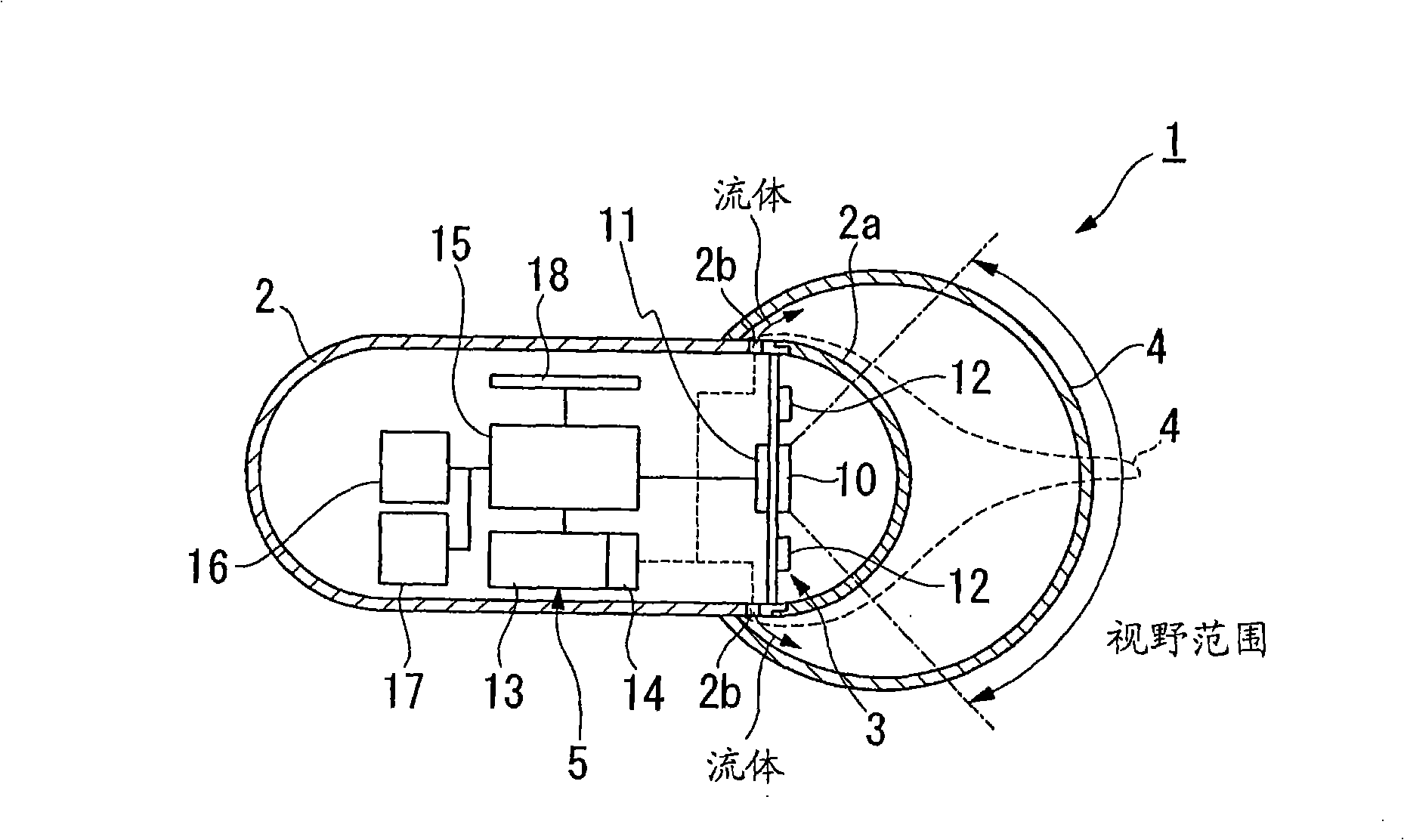
[0116] Refer below figure 1 with 2 A first embodiment of the in-vivo observation device as claimed in the present invention will be described. Such as figure 1 As shown, the in-vivo observation device 1 of this embodiment is provided with a capsule casing 2 for oral intake into the body, an observation system (observation device) 3 installed in the casing 2 for observing the inside of the body, and arranged in the casing 2 so as to cover An optically transparent balloon 4 that is positioned around the periphery of the observation system 3 and can be inflated so as to be in close contact with or attached to body tissue when moving in the body, and by supplying fluid into the balloon 4 to make the balloon 4 Expansion device 5 for expansion.

[0117] In addition, the refractive index n of the transparent material of the balloon 4 a equal to or less than the refractive index n of the fluid supplied to the balloon 4 b (n a ≤n b ).

[0118] The above-mentioned housing 2 is...

no. 2 example

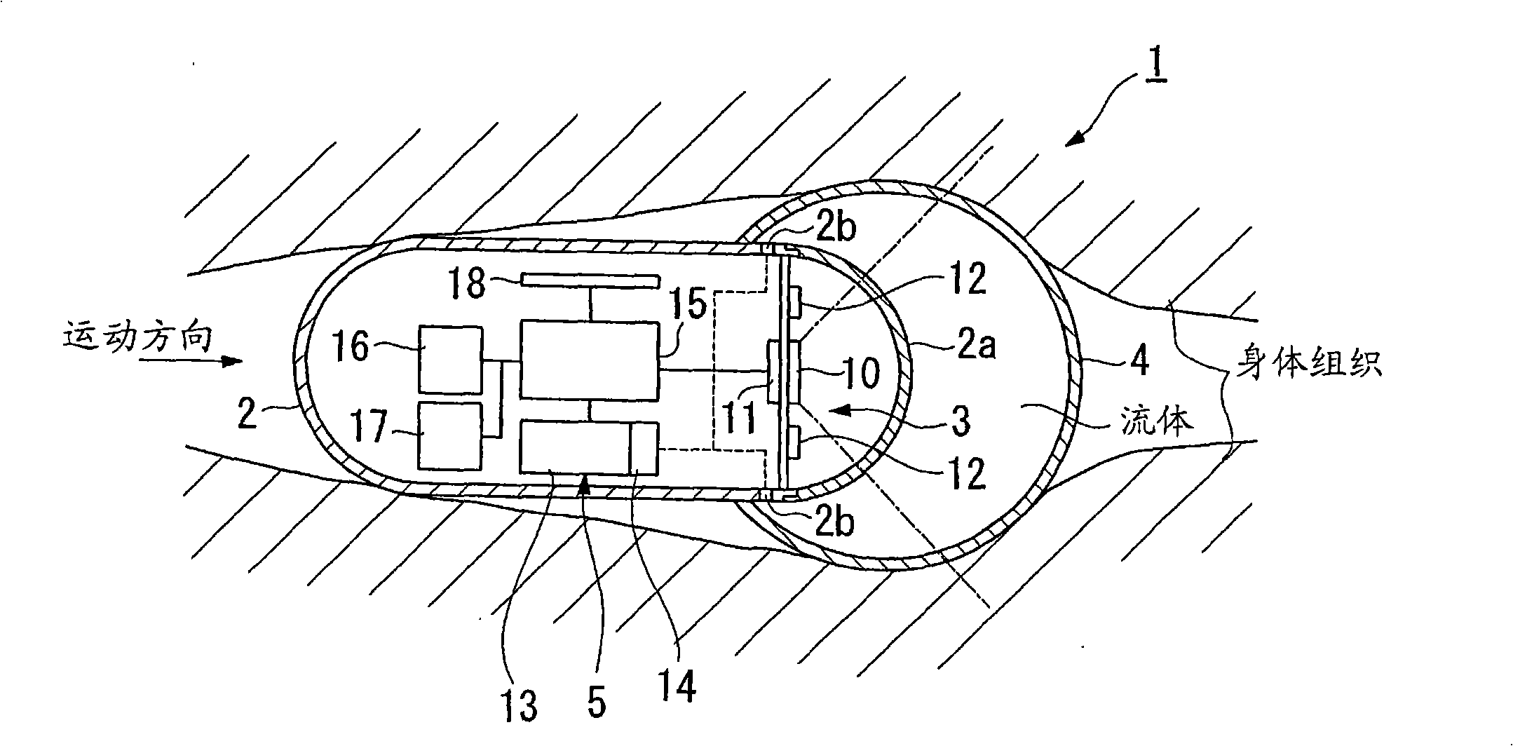
[0133] will then refer to image 3 A second embodiment of the in-vivo observation device as claimed in the present invention will be described.

[0134] In addition, the same constituent features in the second embodiment as those in the first embodiment are denoted by the same reference numerals, and descriptions thereof are omitted.

[0135] The difference between the first embodiment and the second embodiment is that the fluid A contained in the storage portion 13 is supplied to the balloon 4 through the inflation device 5 when the balloon 4 is inflated in the first embodiment. In contrast, the in-vivo observation device 20 of the second embodiment inflates the balloon 4 using bodily fluids in the body.

[0136] That is, if image 3 As shown, the in-vivo observation device 20 of the current embodiment has an acquisition part 22 for acquiring body fluid in the body, and an expansion vessel (an expansion part) for inflating the balloon 4 according to the water content of the...

no. 3 example

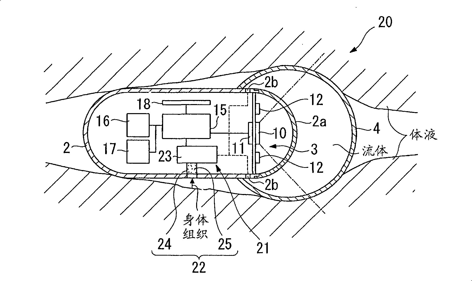
[0143] will then refer to Figure 4 A third embodiment of the in-vivo observation device as claimed in the present invention will be described.

[0144] In addition, the same reference numerals are used to denote those constituent features in the third embodiment that are the same as those in the first embodiment, and descriptions thereof are omitted.

[0145] The difference between the third embodiment and the first embodiment is that, unlike in the first embodiment, the fluid A contained in the storage portion 13 is supplied to the balloon 4 through the inflation device 5 when the balloon 4 is inflated. In contrast, the in-vivo observation device 30 of the third embodiment inflates the balloon 4 using air (fluid) in the body.

[0146] That is, if Figure 4 As shown, the in-vivo observation device 30 of the current embodiment is provided with a pipeline 31 for connecting the outside and inside of the balloon 4 in the housing 2, and the expansion device 32 is provided with a...


PUM


Abstract

This in vivo observation device is equipped with a capsular casing that is taken orally into the body, and an observation device provided within said casing which observes the inside of the body through an observation wall surface of said casing; and, is provided with a contact auxiliary device that causes the observation wall surface to closely contact to body tissue during observation.

Description

[0001] This application is a divisional application of an invention patent application with an application date of October 1, 2004, an application number of 200480005150.6 (PCT / JP2004 / 014488), and an invention title of "in vivo observation device". technical field [0002] The present invention relates to an in-vivo observation device for observing diseased areas such as superficial diseases and the like occurring in the digestive tract and other parts. [0003] This application claims the benefit of Japanese Patent Application No. 2003-343064 filed on October 1, 2003 and Japanese Patent Application No. 2003-361782 filed on October 22, 2003, the contents of which are incorporated herein by reference Reference. Background technique [0004] Inflammatory bowel diseases (IBDs) such as ulcerative colitis and Crohn's disease are diseases of the digestive tract whose causes are still unknown, and there is currently an urgent need to establish various treatment and prevention metho...

Claims


Application Information

Patent Timeline
Patent Type & Authority Applications(China)
IPC IPC(8): A61B1/05A61B1/31A61B5/07A61M31/00A61J3/07
Inventor 河野宏尚内山昭夫泷泽宽伸菊地昭
Owner OLYMPUS CORP
Why Eureka
Try Eureka