
Ultrasonic diagnostic imaging system with spectral and audio tissue doppler

An imaging system and ultrasonic diagnostic technology, applied in the field of medical ultrasonic diagnostic imaging system, can solve the problems of hard to hear, small speakers, distortion, etc., and achieve the effect of improving clarity

Inactive Publication Date: 2008-10-29
KONINKLIJKE PHILIPS ELECTRONICS NV
View PDF1 Cites 9 Cited by
  • Summary
  • Abstract
  • Description
  • Claims
  • Application Information

AI Technical Summary

Problems solved by technology

It is difficult for small speakers to produce very low-pitched sound, and such sound is difficult for most people to hear
Amplifying the volume helps, but can cause distortion
[0009] Conventional FFT periodogram techniques are well suited for weak, broadband blood signals, but tissue Doppler typically has strong, narrowband signals

Method used

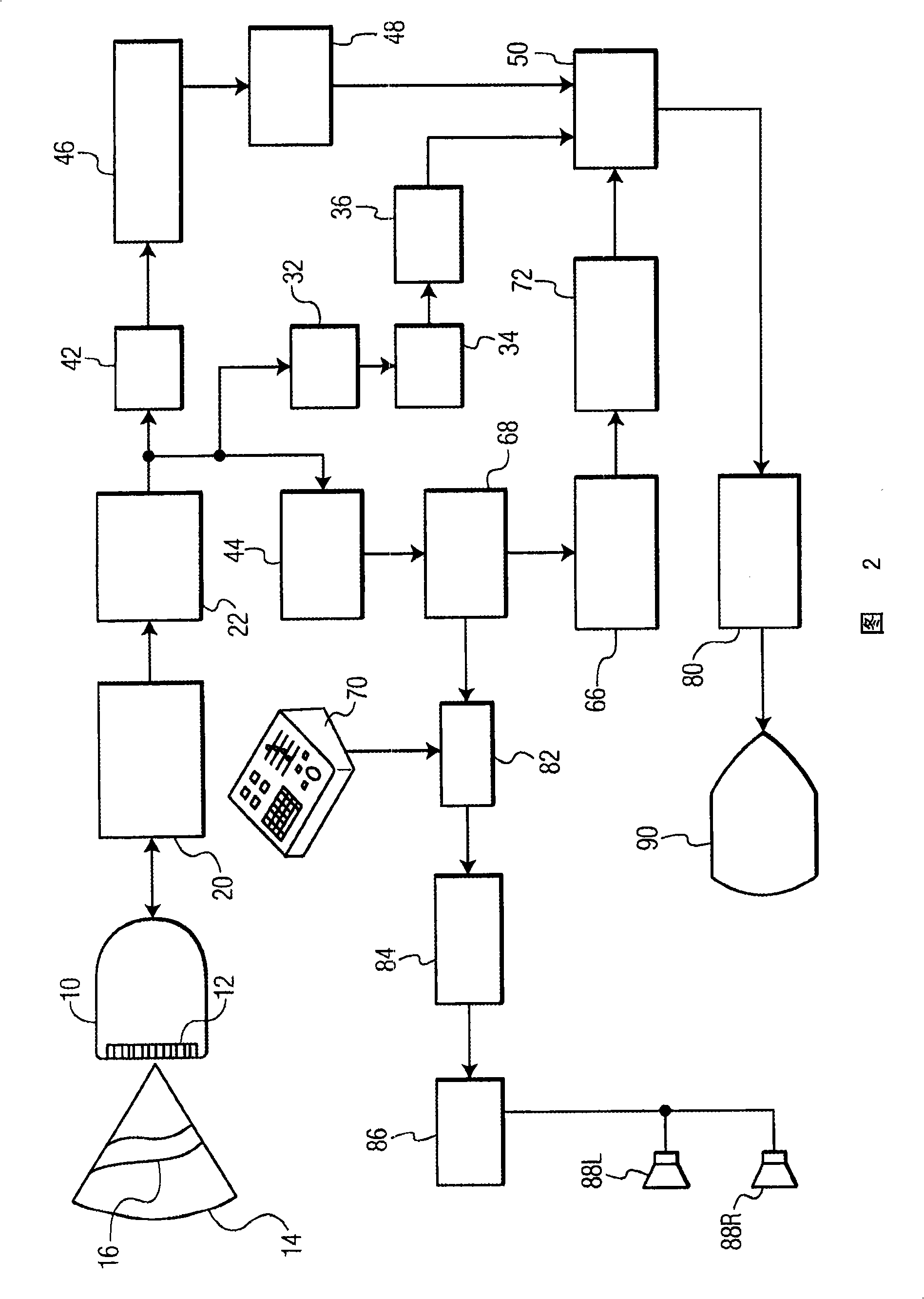
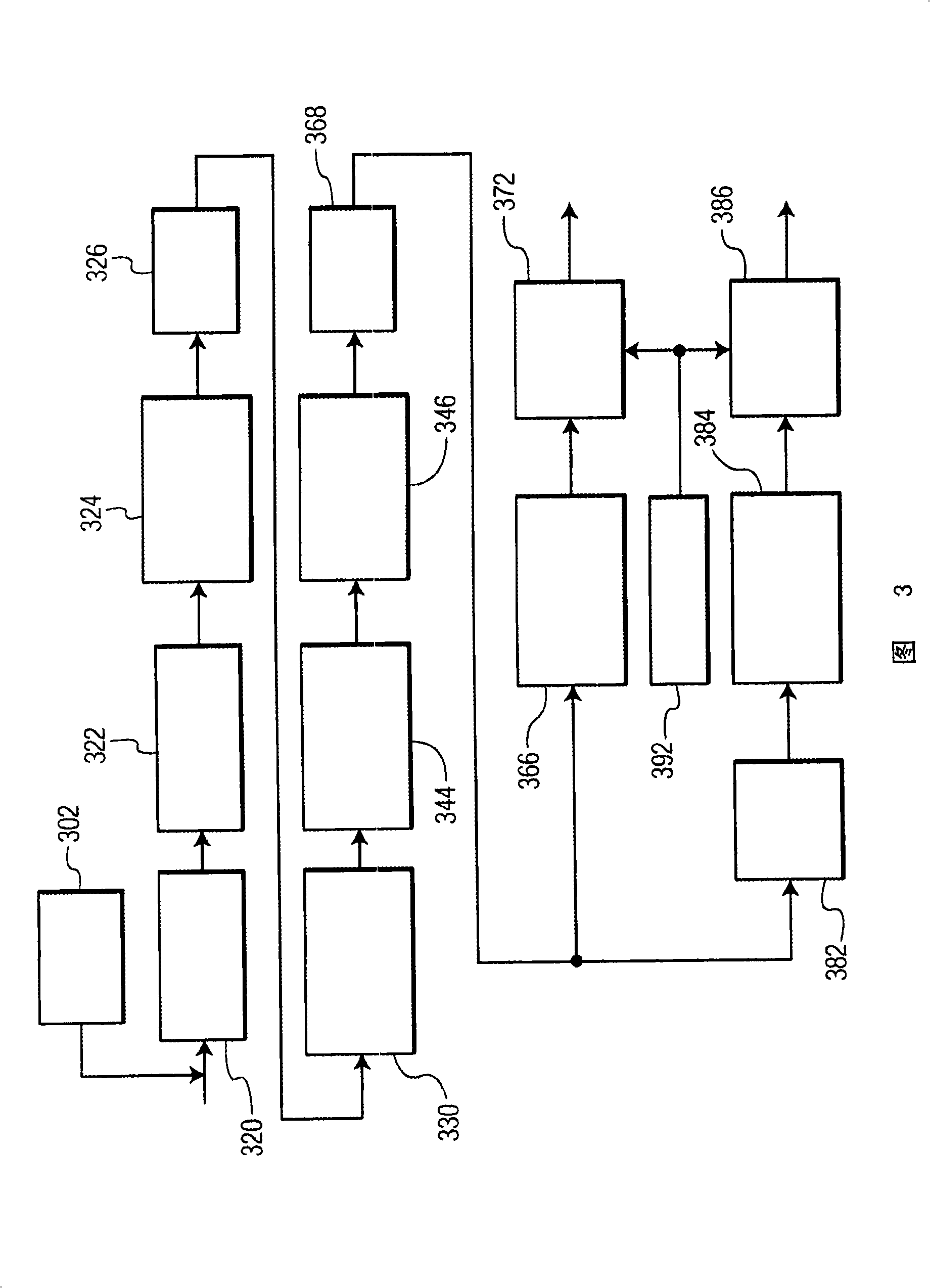

Image

  • Ultrasonic diagnostic imaging system with spectral and audio tissue doppler
  • Ultrasonic diagnostic imaging system with spectral and audio tissue doppler
  • Ultrasonic diagnostic imaging system with spectral and audio tissue doppler

Examples


Embodiment Construction

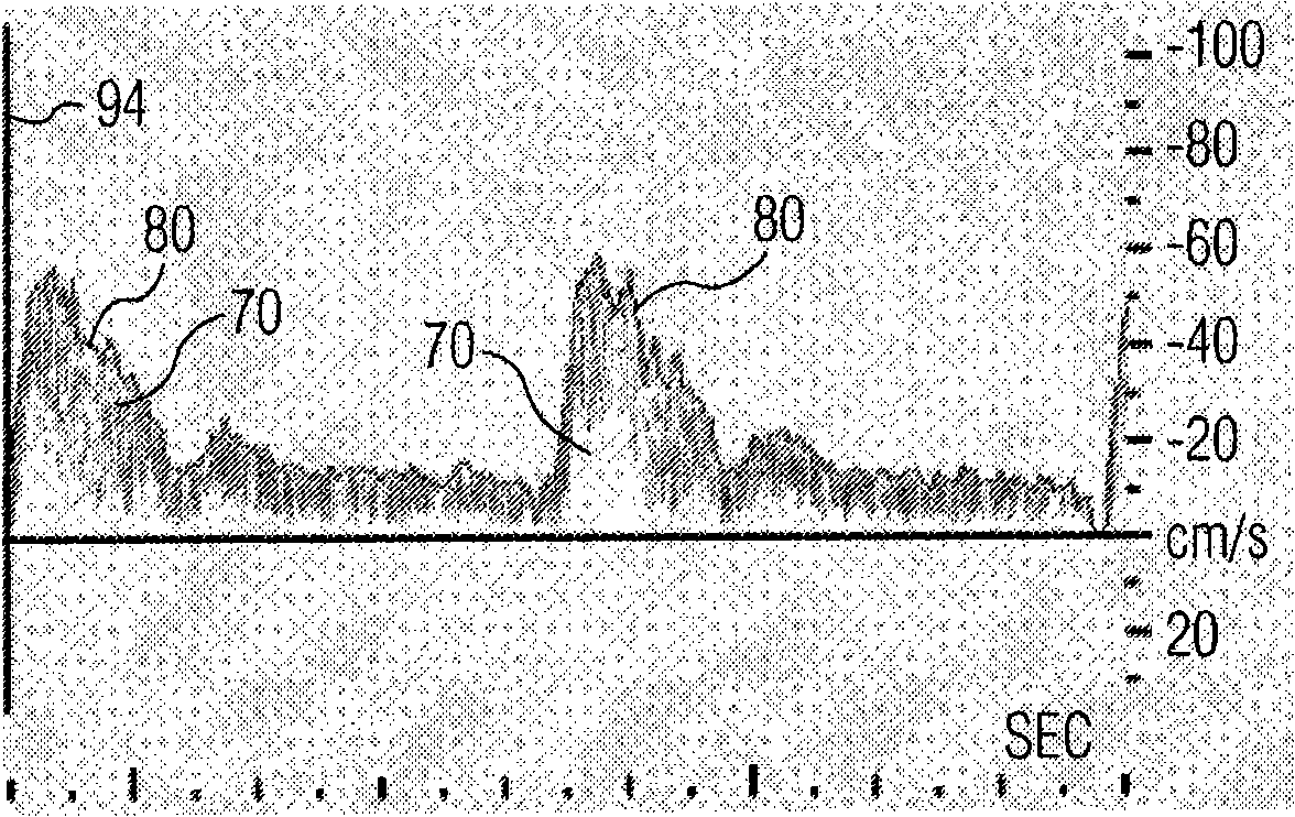
[0016] first reference figure 1 , showing a standard spectral Doppler display of blood flow. The spectrum shows that the scale on the vertical axis 94 is in cm / sec and the scale on the horizontal axis is time (sec). The spectral display is generated by taking a sequence of samples from a certain point (sample volume) in the chamber of the heart or vessel. A group of consecutive samples is called a window and operates as a unit. For example, a spectral Doppler window for blood flow may consist of 128-256 consecutive samples. Sampling within a window is usually weighted, with the greatest weight applied at the center of the window. The weighted samples are then subjected to an FFT, as known in the art, to produce a Fourier series of weighted samples. FFT processing transforms time-domain samples into frequency-domain samples in complex form, with real and imaginary parts. The magnitudes of the samples are calculated and logarithmic values ​​are taken for each magnitude. Ea...


PUM


Abstract

A spectral tissue Doppler processor for an ultrasound system produces Doppler phase shift estimates of sequences of signal samples from a sample volume with a short-lag autocorrelator. The autocorrelation products are summed and an arc tangent taken of each sum to produce angle estimates. The angle estimates, which are proportional to the tissue motion velocity, are plotted, smoothed, and displayed as a spectral tissue Doppler display. The angle estimates are also used to produce the audio Doppler signal which is frequency-adjustable by a user. The spectral Doppler display exhibits good time and velocity resolution for motion which is less than that of blood flow such as myocardial motion. Major causes of blurring, unevenness, and distortion are reduced or eliminated.

Description

technical field [0001] The present invention relates to medical ultrasound diagnostic imaging systems, and more particularly to ultrasound systems that present spectral and audio tissue Doppler information. Background technique [0002] Tissue Doppler ultrasound is used in echocardiology to measure the motion and timing of the heart muscle. An adaptation of ultrasound techniques for analyzing blood velocity is: color flow imaging, and spectral and audio pulsed wave Doppler. In blood flow techniques, clutter filters suppress strong, slow tissue echoes so that very weak, fast echoes from flowing blood can be seen. Tissue Doppler typically does not use a clutter filter because the echoes of interest are strong, slow tissue echoes. In tissue Doppler, what is analyzed is the predominantly slow tissue echo signal, which has a much higher amplitude than the signals from blood, noise, and reverberation. [0003] Spectral Doppler signals for blood flow processing are based on over...

Claims


Application Information

Patent Timeline
Patent Type & Authority Applications(China)
IPC IPC(8): G01S15/89
Inventor D·W·克拉克
Owner KONINKLIJKE PHILIPS ELECTRONICS NV
Learn More