
Tissue separation imaging method based on inversion recovery

A technology of tissue separation and imaging method, which is applied in the direction of using nuclear magnetic resonance imaging system for measurement, magnetic variable measurement, magnetic resonance measurement, etc., can solve the problems of reduced image signal-to-noise ratio, inability to separate images, and reduced signal amplitude

Active Publication Date: 2009-11-04
SIEMENS HEALTHINEERS LTD
View PDF0 Cites 3 Cited by
  • Summary
  • Abstract
  • Description
  • Claims
  • Application Information

AI Technical Summary

Problems solved by technology

[0014] Although the inversion-recovery separation imaging method can ignore the influence of the inhomogeneity of the external magnetic field, and there will be no difficulty in narrow-band pulse design when using the frequency selection method, but when acquiring an image of one of the tissues in the imaging object, Only the echo signals sent by the hydrogen nuclei in this kind of tissue are used, so that the signal amplitude is greatly reduced, and due to the randomness of the noise, the significant reduction of the signal amplitude will reduce the signal-to-noise ratio of the image, thereby affecting the imaging quality
Moreover, this existing inversion recovery separation imaging method cannot calculate and obtain images of various tissues at the same time, nor can it separate images of more than two tissues in the same imaging object.

Method used


Image

  • Tissue separation imaging method based on inversion recovery
  • Tissue separation imaging method based on inversion recovery
  • Tissue separation imaging method based on inversion recovery

Examples


Embodiment Construction

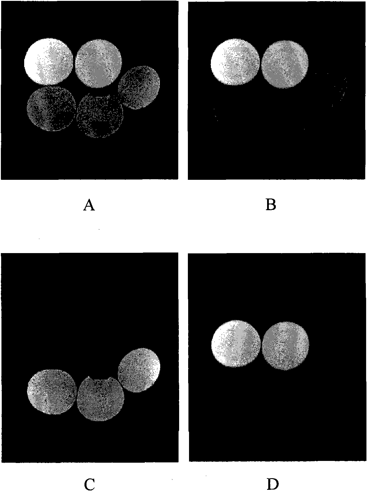
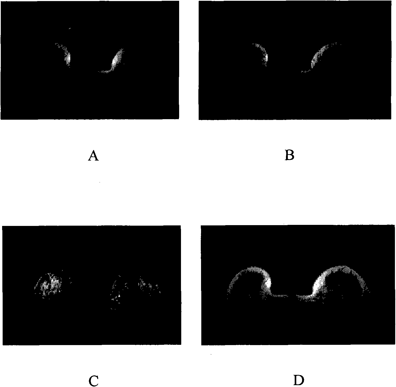
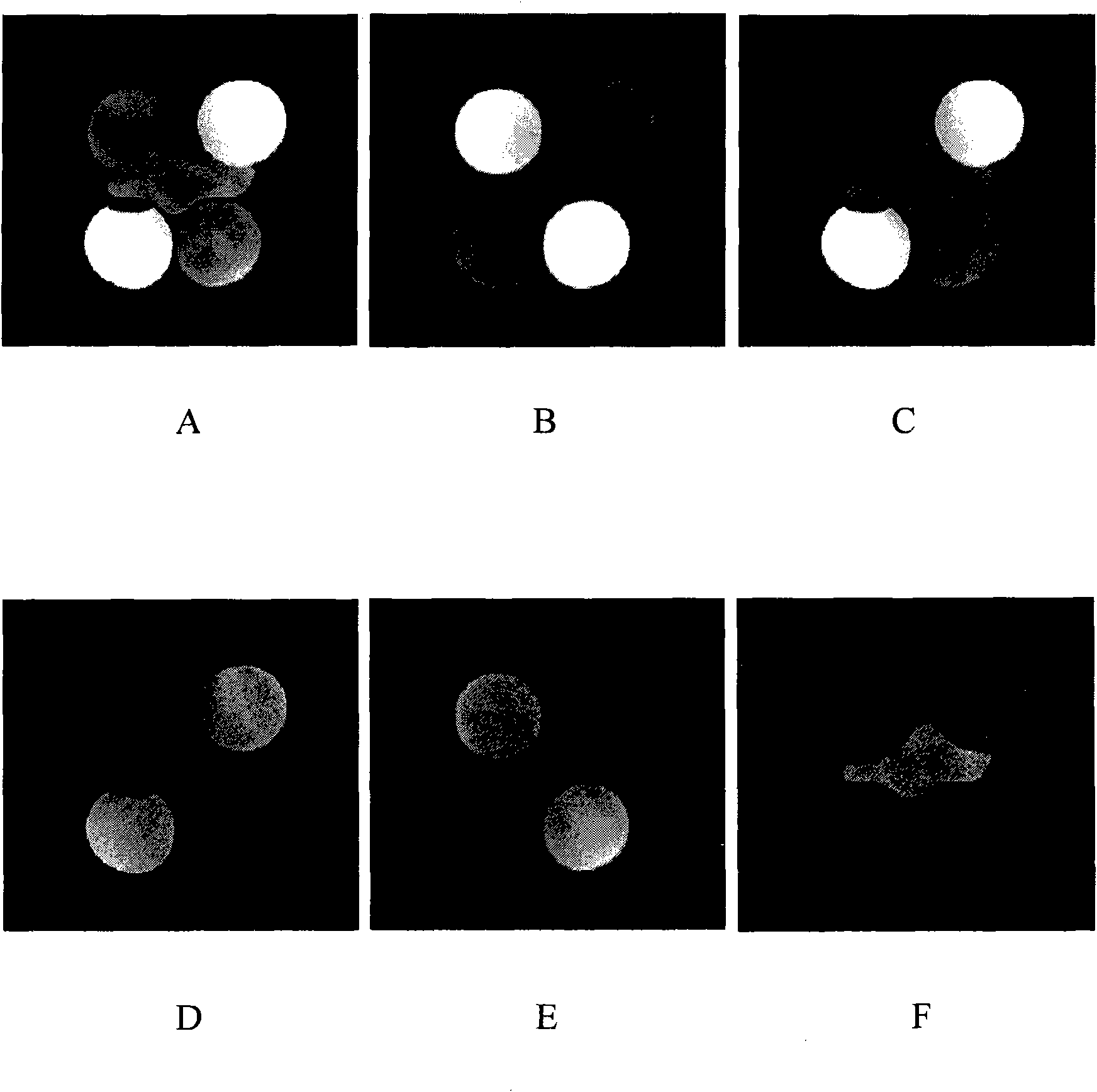
[0035]The difference between the tissue separation imaging method based on inversion recovery of the present invention and the existing inversion recovery tissue separation imaging method mainly lies in that the scanning imaging sequence is used to excite the imaging object, and at different inversion times (Inversion Time; TI; inversion time) The time interval between the 180° pulse and the 90° excitation pulse in the recovery sequence) performs two or more image samplings. The number of sampling times is determined by the number of types of tissues that need to be separated and imaged contained in the imaging object, and generally, the number of sampling times should be equal to or greater than the number of types of tissues. The inversion time TI can be selected arbitrarily. Preferably, the selected principle is to improve the SNR of the sampled image.

[0036] Suppose the image sampled in an inversion time TI is I ∑.TI (x, y), the image is the sum of the images of all kin...


PUM


Abstract

The invention provides a tissue separation imaging method based on inversion recovery, adopting scanning imaging sequence to excite an imaging object, and carrying out image sampling on the imaging object in different inversion times. Each sampling image comprises an image of each tissue, the time of sampling is determined by kind quantity of tissues to be separated and imaged are contained in the imaging object, and a pure tissue image of each tissue is obtained through simultaneous solution according to each sampling image data and proportion relation occupied by each tissue image data in each sampling image. The method is insensitive to the nonuniformity of a magnetic field, obtains the image with higher SNR through the separation imaging, can realize simultaneous separation imaging of multiple tissues, and is widely applied to various low, middle and high magnetic field MR imaging systems.

Description

technical field [0001] The invention relates to magnetic resonance imaging technology, in particular to a tissue separation imaging method based on inversion recovery (IR, Inversion Recovery) technology. Background technique [0002] Magnetic resonance imaging is a technique that uses the phenomenon of magnetic resonance to perform imaging. The principle of magnetic resonance phenomenon mainly includes: atomic nuclei containing a single number of protons, such as hydrogen nuclei widely present in the human body, the protons have spin motion, just like a small magnet, and the spin axes of these small magnets have no certain rules. External magnetic field, these small magnets will be rearranged according to the magnetic force lines of the external magnetic field, specifically in two directions parallel to or antiparallel to the magnetic force lines of the external magnetic field, and the above-mentioned direction parallel to the magnetic force lines of the external magnetic fi...

Claims


Application Information

Patent Timeline
Patent Type & Authority Applications(China)
IPC IPC(8): G01R33/54
Inventor 翁得河
Owner SIEMENS HEALTHINEERS LTD
Learn More