Method for detecting hemostatic capability

A detection method and capability technology, applied in the direction of color/spectral characteristic measurement, etc., can solve problems such as reflecting blood coagulation status and difficulty

Inactive Publication Date: 2009-11-25
XI AN JIAOTONG UNIV
View PDF0 Cites 4 Cited by
  • Summary
  • Abstract
  • Description
  • Claims
  • Application Information

AI Technical Summary

Problems solved by technology

In fact, this method is difficult to truly or comprehensively reflect the coagulation status

Method used


Image

  • Method for detecting hemostatic capability
  • Method for detecting hemostatic capability
  • Method for detecting hemostatic capability

Examples


Embodiment 1

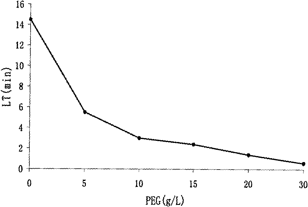
[0018] Example 1: Select 0.1mol / L Tris buffer as the buffer system (pH 7.35), appropriately screened coagulation activators, appropriate amount of calcium chloride as a coagulant accelerator, appropriate amount of Tween-80 as a stabilizer, and add different concentrations of PEG -6000 (5 ~ 30g / L) to adjust the reaction lag time.

[0019] figure 1 It shows the effect of different concentrations of polyethylene glycol on the OHP lag time, and the coagulation reaction is too fast or too slow, which will affect the sensitivity of the test. According to calculations, the appropriate coagulation reaction lag time should be within the range of 2 to 3 minutes. Therefore, a certain amount of polyethylene glycol-6000 (PEG-6000) was added to the OHP test system to adjust the lag time (lag time, LT) of the blood coagulation reaction and improve the sensitivity of the test. According to the screening results of different concentrations of PEG-60000, the concentration of PEG-6000 to keep ...

Embodiment 2

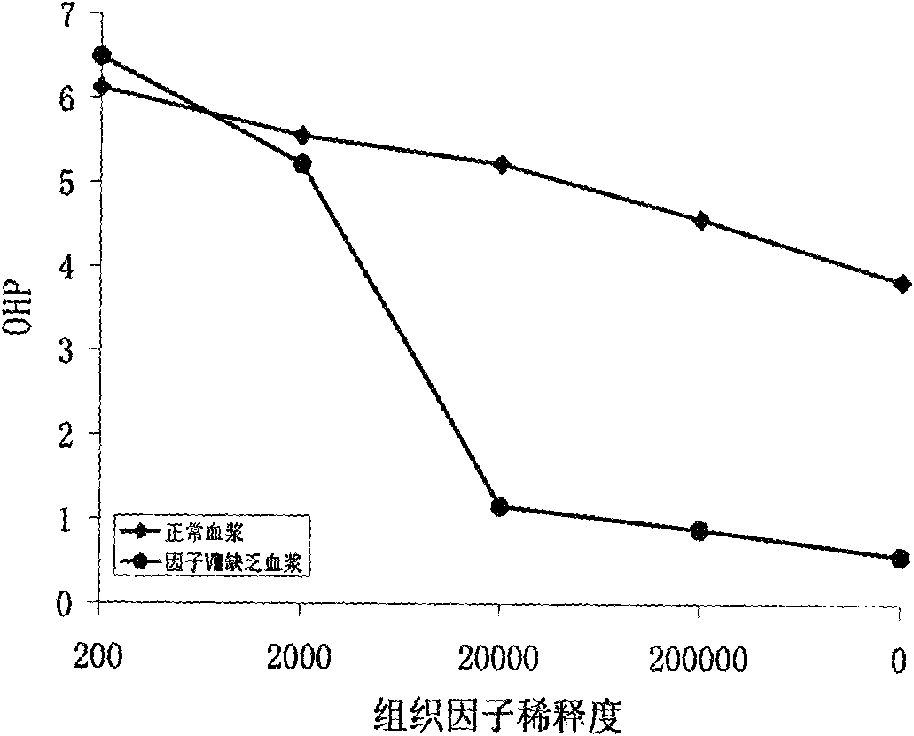
[0020] Embodiment 2: Select 0.1mol / L Tris buffer as the buffer system (pH 7.35), PEG-6000 20g / L, Tween-80 10g / L, CaCl 2 5mmol / L, tissue factor (TF) concentration from 0 to 1 / 200000, measure normal plasma and factor VIII lack of plasma coagulation curve.

[0021] figure 2 Show the effect of different concentrations of TF on OHP, OHP test requires full activation of coagulation reaction, but the usual coagulation activators do not meet this requirement. In the presence of relatively high concentrations of TF, there was no significant difference in OHP values ​​between normal control plasma and factor VIII-deficient plasma. However, when the TF concentration fell below a 1 / 20000 dilution, the OHP values ​​were significantly lower in Factor VIII-deficient plasma compared to control plasma. It shows that low concentration of TF can activate both intrinsic and extrinsic blood coagulation pathways at the same time. Therefore, a 1 / 20000 dilution of TF was selected as a coagulatio...

Embodiment 3

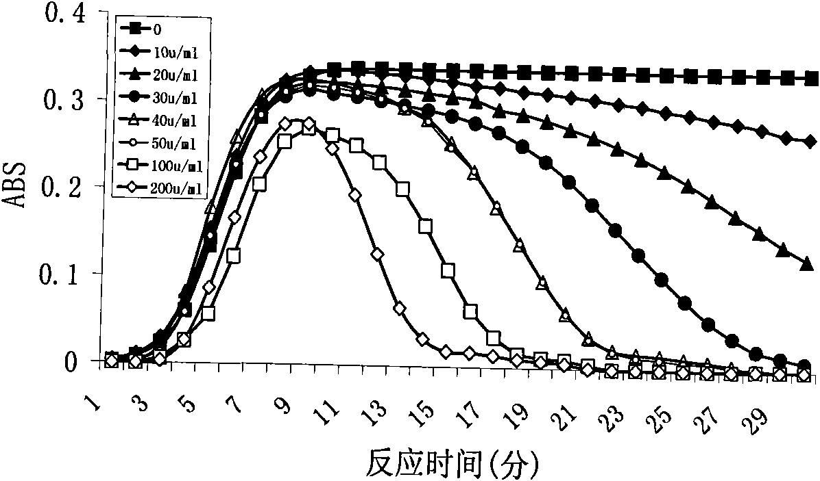
[0022] Embodiment 3: Select 0.1mol / L Tris buffer as the buffer system (pH 7.35), PEG-6000 20g / L, Tween-80 10g / L, CaCl 2 5mmol / L, tissue factor (TF) concentration 1 / 20000, UK concentration from 10U to 200U / ml.

[0023] image 3 To show the influence of different concentrations of UK on OHP, plasminogen activator (UK) was added to the experimental system to activate the fibrinolytic system to measure the fibrinolytic ability. From image 3 It can be clearly seen that when the UK concentration is less than 30U / ml, the generated fibrin cannot be effectively degraded; when the UK concentration is greater than 50U / ml, although the fibrin can be rapidly degraded, the formation of fibrin is significantly reduced. Therefore, we choose the UK concentration of 40U / ml.


PUM


Abstract

The invention discloses a method for detecting hemostatic capability. The method comprises: preparing anticoagulant blood from venous blood and anticoagulant with 0.13mol/L of sodium citrate in a volume ratio of 9:1; centrifuging an anticoagulant blood sample for 15 minutes at a speed of 3,000 r/min and sucking plasma to be detected; performing OHP determination of overall hemostatic capability, OCP determination of overall blood coagulation capability and OFP judgment of overall fibrinolytic capability; indicating the existence of blood coagulation disturbance when OCP and OHP are less than the lower limit of normal reference values; indicating the existence of hypercoagulable state when the OCP and the OHP are greater than the upper limit of the normal reference values; and indicating the reduction of fibrinolytic function when OFP value is less than 41.61 percent. The method has the advantages of good stability and repeatability, easy operation and low cost, and is worthy of popularization.

Description

technical field [0001] The invention relates to an experimental method for hemostasis and thrombus detection, in particular to a detection method for blood plasma hemostatic ability. Background technique [0002] For a long time, people have been trying to establish a test that can reflect the overall hemostatic ability and be used to evaluate the body's hemostatic balance. In fact, as early as 1995, Hemker et al. established the so-called endogenous thrombin potential (ETP) assay method for evaluating the coagulation balance of the body. This method activates plasma with a mixture of low concentration TF and phospholipids, uses artificial chromogenic substrates to measure thrombin generation, and calculates ETP according to the thrombin generation diagram (Thrombogram). Increased ETP is seen in cases of prothrombotic and / or hypercoagulable states, including antithrombin deficiency, high expression of prothrombin (prothrombin A20210C), protein C / protein S deficiency, factor...

Claims


Application Information

Patent Timeline
Patent Type & Authority Applications(China)
IPC IPC(8): G01N21/31
Inventor 吴锋马西李晓会庄贵华贺林车金
Owner XI AN JIAOTONG UNIV
Why Eureka
Try Eureka