Three-dimensional medical image segmentation method

A technology of medical images and three-dimensional images, which is applied in image analysis, image data processing, instruments, etc., can solve problems such as the difficulty in determining the precise threshold, the inability to accurately distinguish the characteristics of different regions, and the over-segmentation or under-segmentation of multiple regions. Achieve the effects of strong adaptability to morphological separation and merging, clear and reliable image local features, and less data storage space requirements

Active Publication Date: 2010-09-01
四川锦江电子医疗器械科技股份有限公司
View PDF7 Cites 33 Cited by
  • Summary
  • Abstract
  • Description
  • Claims
  • Application Information

AI Technical Summary

Problems solved by technology

[0005] 1) It is difficult to determine the precise threshold value, and the difference of medical images leads to the difference of threshold value;
[0006] 2) In the case that the gray scales of multiple regions of the image are roughly the same, the characteristics of different regions cannot be accurately distinguished, and the segmentation effect cannot be achieved;
[0007] 3) In the case of obvious differences in the gray scales of multiple regions of the image, the single gray threshold can reduce the discrimination effect of different regions, and even damage the image quality of the segmented region
[0009] 1) The region growing method can only be segmented for a single region or multiple regions without connectivity
In the case of multi-regions with certain connectivity, the region growing method cannot distinguish the connected edges of multi-regions, and it cannot achieve the purpose of multi-region segmentation;
[0010] 2) The region growing method will cause inaccurate image segmentation and extraction when the image gray level is uneven and the difference is large.
[0012] 1) The calculation of energy characteristics is easy to fall into local minimization;
[0013] 2) The segmentation of the image region of interest with large shape changes is not ideal, and there is an over-segmentation phenomenon;
[0014] 3) When the shape of the region of interest in the sequence image is separate

Method used


Image

  • Three-dimensional medical image segmentation method

Examples


Embodiment Construction

[0045] All features disclosed in this specification, or steps in all methods or processes disclosed, may be combined in any manner, except for mutually exclusive features and / or steps.

[0046] Any feature disclosed in this specification (including any appended claims, abstract and drawings), unless expressly stated otherwise, may be replaced by alternative features which are equivalent or serve a similar purpose. That is, unless expressly stated otherwise, each feature is one example only of a series of equivalent or similar features.

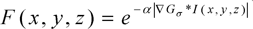
[0047] A three-dimensional medical image segmentation method, comprising the steps of:

[0048] In the first step, in the case of interactive operation, the CT / MRI sequence 2D medical image is reconstructed according to the image sequence and the input gray threshold, and a 3D image containing all regions of interest is generated that meets the gray threshold:

[0049] During the generation process, the two-dimensional image can be sampled an...


PUM


Abstract

The invention discloses a three-dimensional medical image segmentation method. The method comprises the following steps: firstly performing three-dimensional superposition reconstruction on sequential two-dimensional medical image, and then performing Gaussian filter smoothing in a three-dimensional space; carrying out three-dimensional space contraction computation on the three-dimensional medical image, and calculating and recording the contraction orders and the energy characteristics of every three-dimensional pixel point; performing three-dimensional space extension on the three-dimensional medical image in a plurality of areas by a level set method; and finally extracting and segmenting the three-dimensional pixel points in the three-dimensional image according to information of interested areas, and respectively generating a plurality of three-dimensional images of the interested areas. The method has positive effects of strong timeliness, high computing efficiency, high segmentation accuracy, and clear and reliable image local features, and can achieve simultaneous segmentation of a plurality of areas.

Description

technical field [0001] The invention relates to a method for segmenting a three-dimensional medical image, in particular to a method for segmenting a three-dimensional medical image based on a multi-region contraction-expansion collision strategy. Background technique [0002] In the medical field, 3D medical image segmentation is used to segment the region of interest or lesion in the 3D medical image, to observe and analyze the morphology, characteristics and other pathological conditions of the region of interest or lesion, and to conduct 3D medical Image reconstruction and fusion, etc. Especially for medical images of the heart, it is necessary to segment and extract multiple regions of the medical image of the heart at the same time, such as the segmentation of left and right atria or left and right ventricles. [0003] Generally speaking, most medical image segmentation methods are basically based on CT / MRI sequence images to perform image segmentation in two-dimensio...

Claims


Application Information

Patent Timeline
IPC IPC(8): G06T7/00G06T17/00
Inventor 李楚文杨勇史天才
Owner 四川锦江电子医疗器械科技股份有限公司
Why Eureka
Try Eureka