Cell separation method based on morphology

A technology based on morphology and separation methods, applied in the fields of computing, computer vision, image processing, and biomedicine, can solve problems that affect the calculation of cell morphology parameters and cannot effectively separate cells that adhere

Inactive Publication Date: 2010-10-06
ZHEJIANG UNIV OF TECH
View PDF2 Cites 20 Cited by
  • Summary
  • Abstract
  • Description
  • Claims
  • Application Information

AI Technical Summary

Problems solved by technology

[0004] In order to overcome the shortcomings of the existing cell separation methods that cannot effectively separate the adhered cells and a

Method used


Image

  • Cell separation method based on morphology
  • Cell separation method based on morphology
  • Cell separation method based on morphology

Examples


Embodiment Construction

[0049] The present invention will be further described below in conjunction with the accompanying drawings.

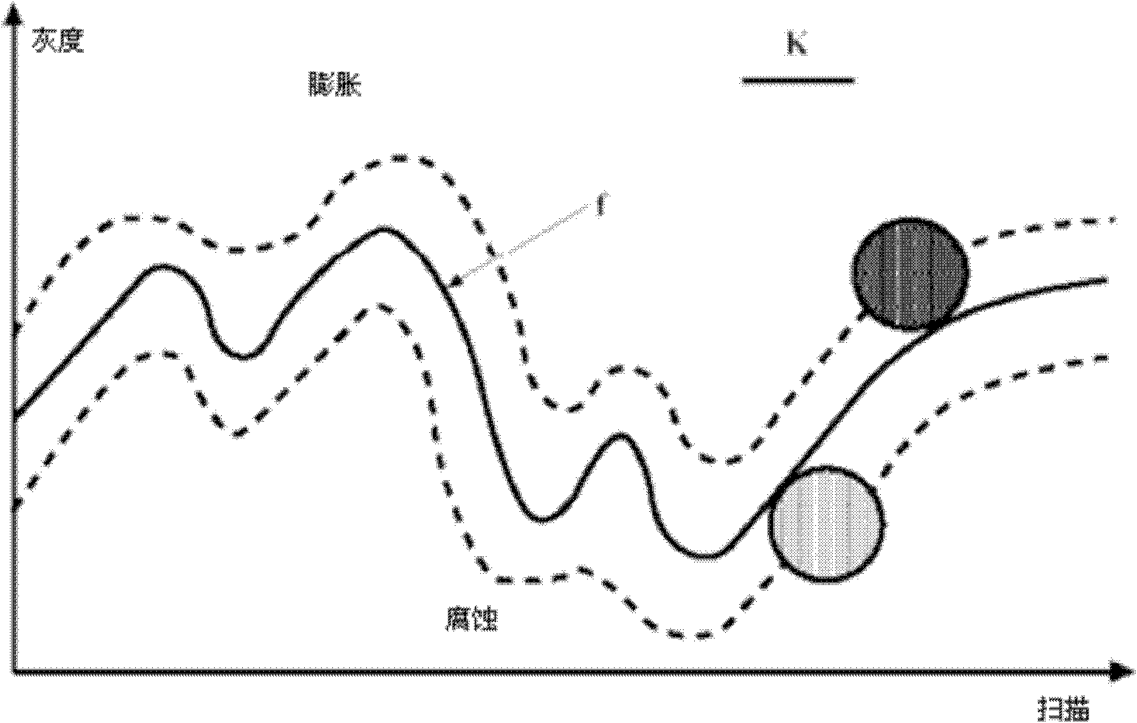
[0050] refer to Figure 1 to Figure 11 , a cell separation method based on morphology, the cell separation method comprises the following steps:

[0051] 1), area mark:

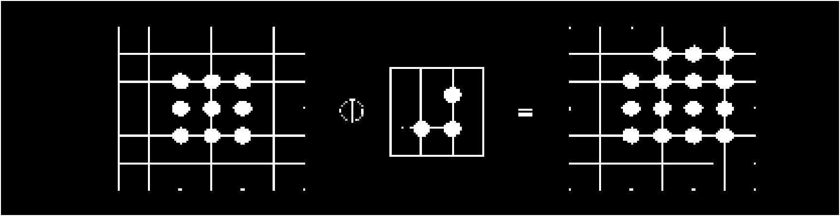
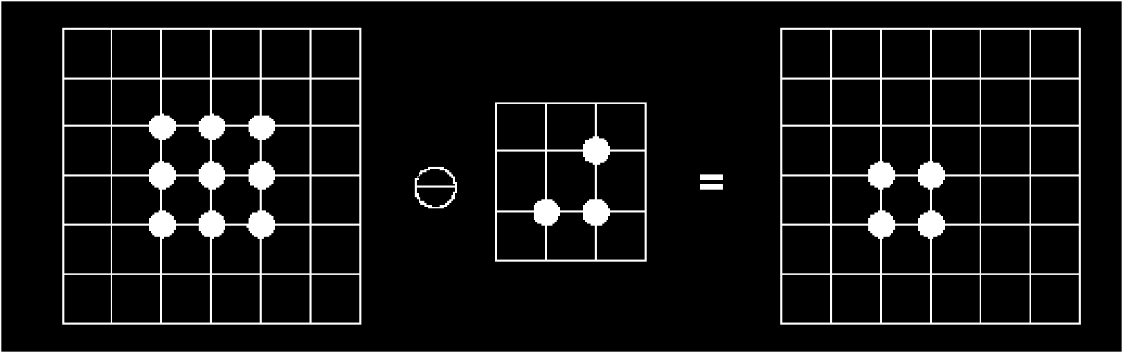
[0052] For the binary image of the cell image segmentation result, the morphological markers are as follows:

[0053] e) Custom structural elements, including shape and size;

[0054] f) use the structural element to corrode the binary image to obtain the internal representative area;

[0055] g) Morphological finishing operation and filling method to eliminate small noise areas, and construct a new binary image containing internal representative areas;

[0056] h) Obtain the marker values ​​of each region with the watershed algorithm on the new binary map;

[0057] Obtain the mark value of each adhesion area through the above marks;

[0058] 2), extract single cells:

[0059] 2.1), the process...


PUM


Abstract

The invention relates to a cell separation method based on morphology, which comprises the following steps that: (1) region labeling: a binary image of a result is cut for a cell image, and the label values of all adhesion areas are obtained by labeling based on norphological labeling, and (2) a monomer cell is extracted; and 2. (1) a process of region extraction, (2) first, a label map with adhesion areas are found out from a regional label map, a circular structure with the structural element of 1 is selected and expanded until the boundary of the label map exceeds the outer boundary of an adhesion map, and the size of the label map is the whole of one monomer cell in adhesion cells, and (3) a reconstructed new binary image set is traversed, and the attribution of all monomers are determined according to coordinate and area information, and an image set is established. The cell separation method based on morphology can effectively separate adhered cells and prepares for accurately calculating the morphological parameters of the cells.

Description

technical field [0001] The invention relates to image processing, biomedicine, computer vision and calculation methods, especially a single cell separation method for cell image segmentation. Background technique [0002] In the medical field, it is usually necessary to segment cells from cell images. Due to the non-homogeneity of cell images and the overlap of spatial positions, there are overlapping images of many cells and some over-segmented regions in the nucleus in the segmentation results. If the shape is directly calculated without processing, it is a challenge for the partial attribution of over-segmentation and the accuracy of shape parameter calculation. [0003] Defects in the traditional cell separation method: 1. It cannot effectively separate the adhered cells, and cannot extract single cells from the adhered cells; 2. It affects the accuracy of the calculation of the morphological parameters of the cells. Contents of the invention [0004] In order to over...

Claims


Application Information

Patent Timeline
IPC IPC(8): G06T5/00C12N5/00
Inventor 陈胜勇赵明珠陈敏姚春燕张厚祥张建伟
Owner ZHEJIANG UNIV OF TECH
Why Eureka
Try Eureka