

Magnetic resonance imaging apparatus
A technology of magnetic resonance imaging and magnetic resonance signals, which is used in measurement devices, measurement of magnetic variables, medical science, etc.
- Summary
- Abstract
- Description
- Claims
- Application Information
AI Technical Summary
Problems solved by technology
Method used
Image

Examples
Embodiment Construction
[0036]Hereinafter, an embodiment of the present invention will be described with reference to the drawings.
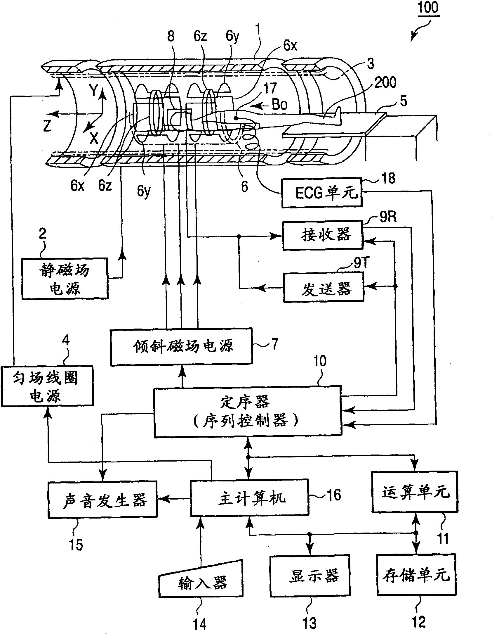
[0037] figure 1 It is a diagram showing a schematic configuration of a magnetic resonance imaging apparatus (MRI apparatus) 100 according to the present embodiment.
[0038] This MRI apparatus 100 includes a bed unit, a static magnetic field generating unit, a gradient magnetic field generating unit, a transmitting and receiving unit, and a control / calculation unit. The bed moves the placed subject 200 . The static magnetic field generator generates a static magnetic field. The gradient magnetic field generator generates a gradient magnetic field for adding positional information to the static magnetic field. The transmitting and receiving unit transmits and receives radio frequency signals. The control computing unit is in charge of overall system control and image reconstruction. In addition, the MRI apparatus 100 includes a magnet 1, a static magnetic field pow...
PUM
 Abstract
Description
Claims
Application Information