Virtual microscope system for monitoring the progress of corneal ablative surgery and associated methods

A technology of corneal surgery, stereo microscope, applied in the field of surgery, which can solve the problems of patient discomfort, damage to workflow, etc.

Inactive Publication Date: 2010-12-01
ALCON REFRACTIVEHORIZONS
View PDF0 Cites 17 Cited by
  • Summary
  • Abstract
  • Description
  • Claims
  • Application Information

AI Technical Summary

Problems solved by technology

[0005] However, illuminating the eye with such illumination to detect flap position, debris, and hydration is uncomfortable for the patient, and using a slit lamp to detect flap repositioning and general eye condition impairs workflow
Also, white light may not provide optimal enhancement of all parts of the eye for visualization

Method used


Image

  • Virtual microscope system for monitoring the progress of corneal ablative surgery and associated methods
  • Virtual microscope system for monitoring the progress of corneal ablative surgery and associated methods
  • Virtual microscope system for monitoring the progress of corneal ablative surgery and associated methods

Examples


Embodiment Construction

[0018] will now refer to Figure 1-7 A description is given of preferred embodiments of the present invention.

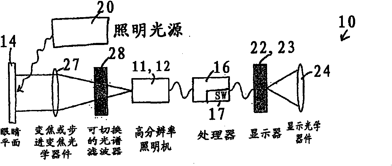
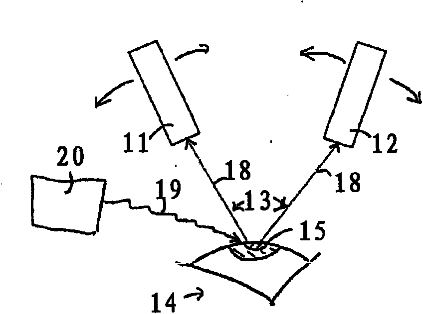
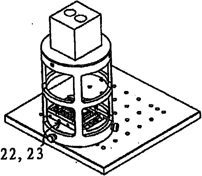
[0019] figure 1 The system schematic diagram of FIG. 2 shows the elements of an exemplary embodiment of the system 10 of the present invention for allowing a surgeon to monitor a corneal surgical procedure. The system 10 includes a first high-resolution color camera 11 and a second high-resolution color camera 12 ( figure 2 ), in a specific embodiment, the first high-resolution color camera 11 and the second high-resolution color camera 12 are adjustable in angular separation 13, and can focus on a part of the eye 14 (for example, the cornea 15) superior. An exemplary surgical procedure to which the system 10 may be applied is LASIK surgery, but should not be limited thereto, and may also apply to pupillometry, other eye measurements such as corneal birefringence, and may also be applied to Other ophthalmic procedures where an operating microscope may be used, ...


PUM


Abstract

A system for visualizing an eye of a patient during corneal surgery includes a processor and a first and second camera in signal communication with the processor. The cameras are positionable for focusing on a cornea positioned for surgery. A first and a second display and optics therefor are in signal communication with the processor and are positionable for viewing through a first and a second eyepiece of a stereo microscope, respectively. Software is resident on the processor for receiving a first and second corneal image from the first and second cameras, for processing the received first and second images for display, and for transmitting the processed first and second images to the first and the second displays, respectively, via the display optics. The displays can then be viewed by a surgeon through the microscope at least during the surgery.

Description

technical field [0001] This application claims priority to US Provisional Application No. 60 / 015,853, filed December 21, 2007. [0002] The present invention relates generally to surgical methods, and in particular to systems and methods for monitoring the progress of corneal ablative surgery. Background technique [0003] Performing LASIK (laser in situ keratomileusis, laser in situ keratomileusis) surgery typically involves excision of a thin flap in the cornea, which is then lifted and folded back along the hinge, so that it The underlying corneal stroma is exposed. Refractive surgery is performed using an ablating laser and the flap is replaced. [0004] Several methods have been used to avoid or detect any "wrinkle", or striae, in the corneal flap after repositioning on the stroma. For example, the cornea can be marked prior to cutting so that the marks can be used to realign the flap. Another method is to use a surgical (direct-view) microscope and a diffuse, broad...

Claims


Application Information

Patent Timeline
Patent Type & Authority Applications(China)
IPC IPC(8): A61B3/13G02B21/22A61F9/007G02B21/06G02B21/00G02B21/36A61F9/01
CPCG02B21/0012A61B3/132G02B21/22A61F9/00804A61F2009/00872
Inventor R·利贝兰克
Owner ALCON REFRACTIVEHORIZONS
Why Eureka
Try Eureka