Countertop ultrasound imaging device and method of using the same for pathology specimen evaluation

An imaging device and ultrasonic imaging technology, applied in ultrasonic/sonic/infrasound image/data processing, re-radiation of sound, ultrasonic/sonic/infrasound diagnosis, etc., can solve problems such as easy to miss

Active Publication Date: 2011-04-06
罗伯特·E·山德斯通
View PDF4 Cites 4 Cited by
  • Summary
  • Abstract
  • Description
  • Claims
  • Application Information

AI Technical Summary

Problems solved by technology

For example, lymph node tumor metastases can be so subtle that they can be easily missed even with several cuts through the tissue sample containing the lymph nodes
Depending on the purpose of tissue sample examination, each microscope slide prepared can take 5-20 minutes of the technician's time

Method used


Image

  • Countertop ultrasound imaging device and method of using the same for pathology specimen evaluation
  • Countertop ultrasound imaging device and method of using the same for pathology specimen evaluation
  • Countertop ultrasound imaging device and method of using the same for pathology specimen evaluation

Examples


Embodiment Construction

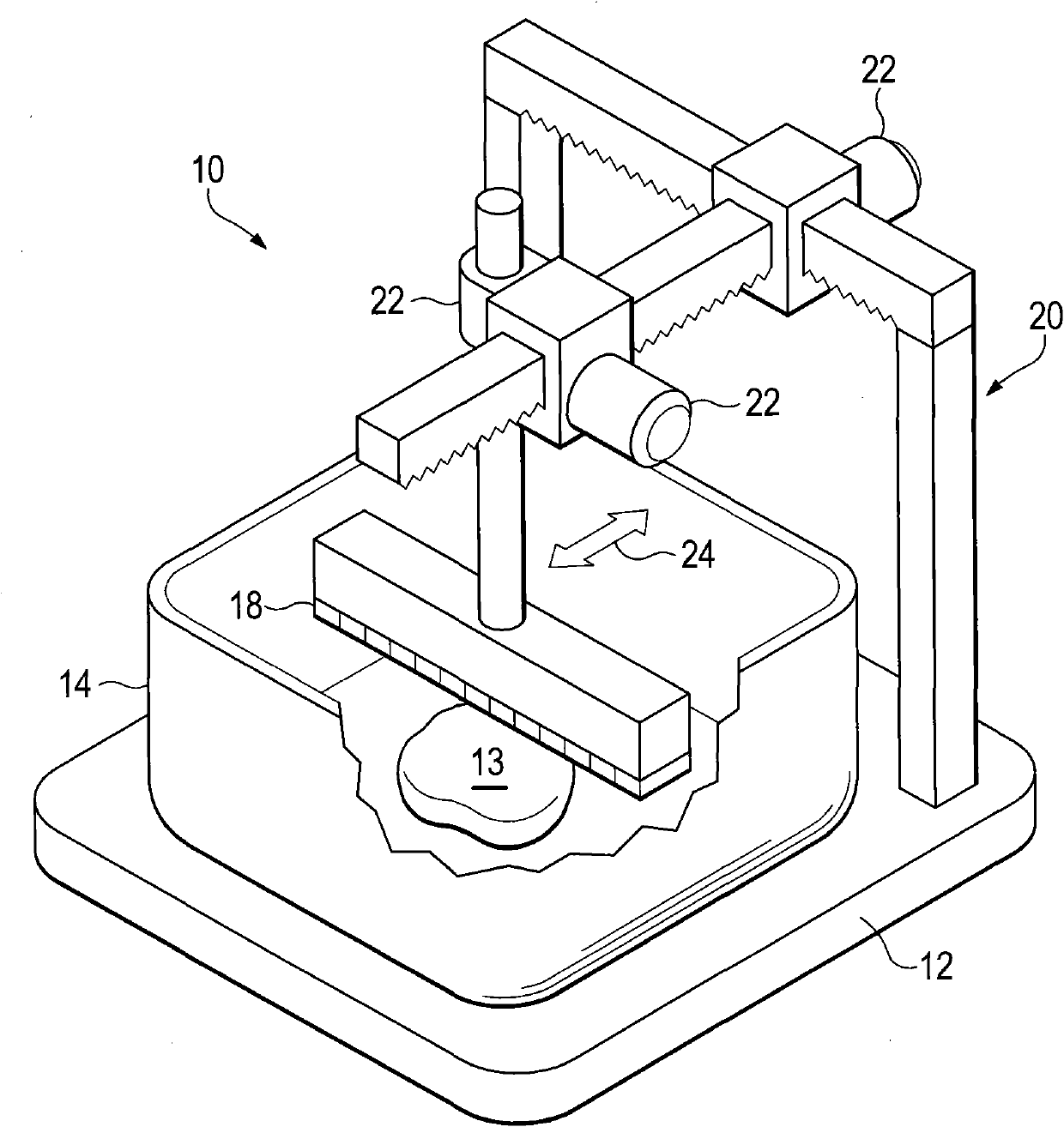
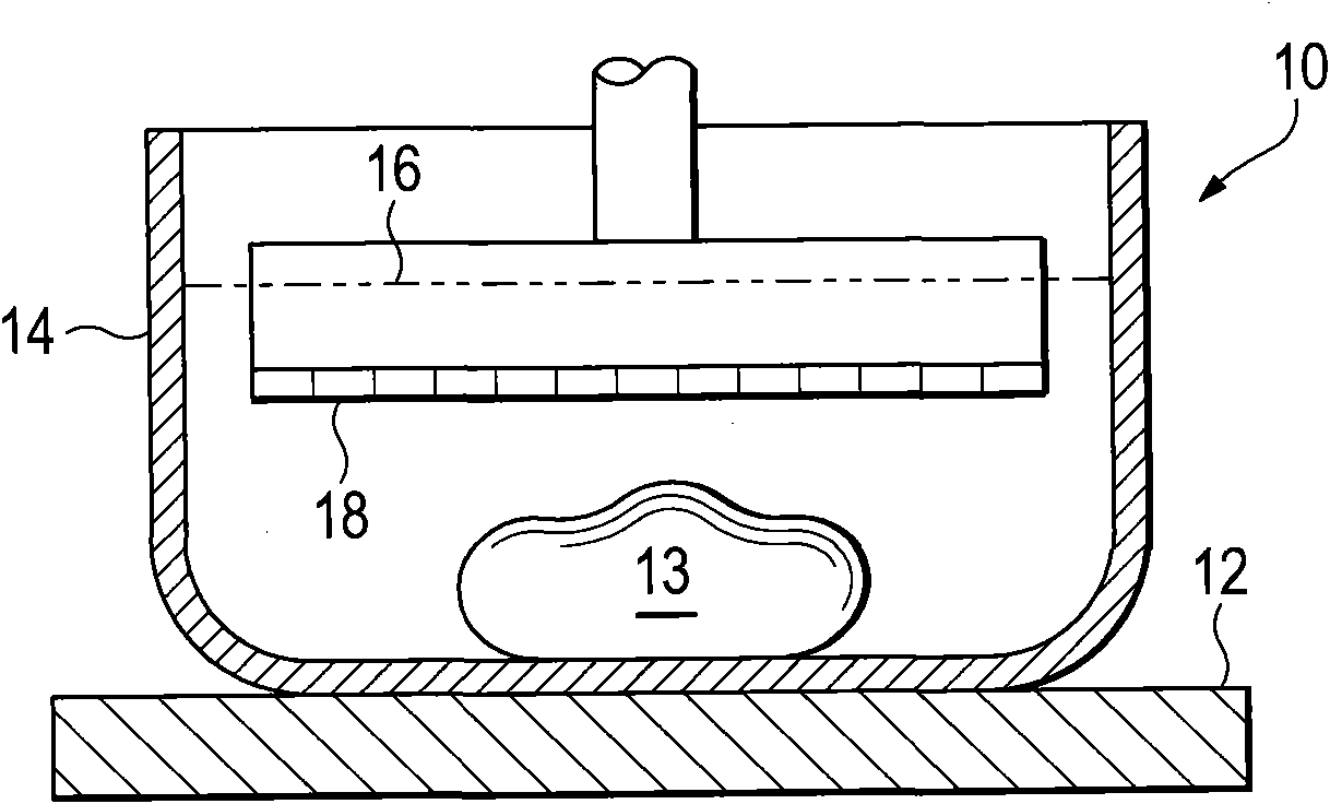
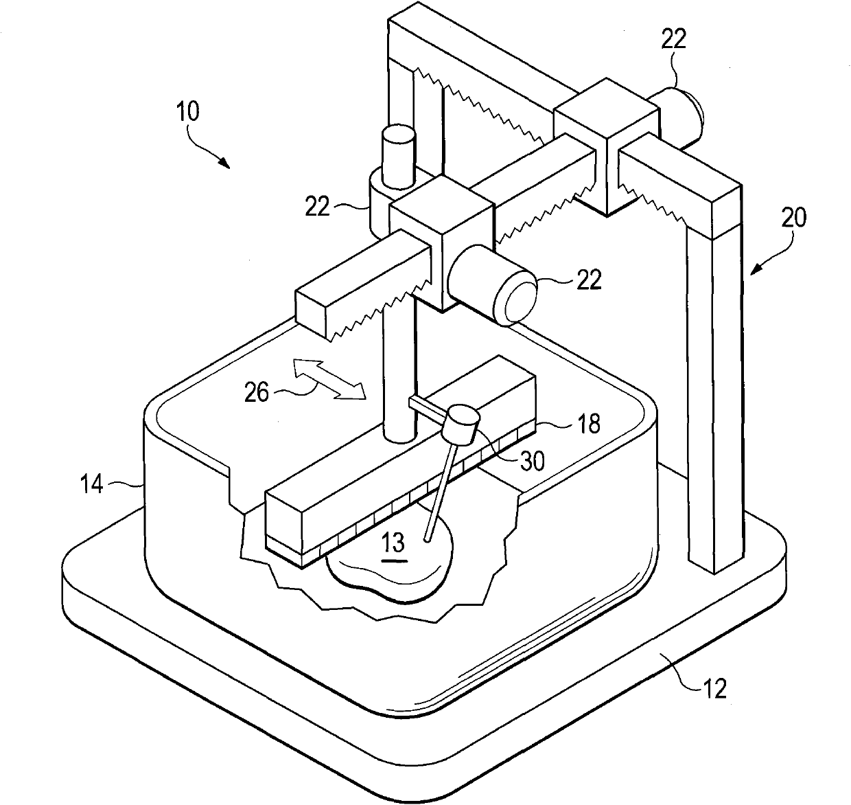
[0016] A preferred embodiment of the present invention is an ultrasound imaging apparatus 10 that can be conveniently supported by a flat surface, such as a laboratory bench, that can receive and image a tissue sample. The device comprises a base 12 supporting a container 14 within which a sample 13 can be placed and which can be filled with a saline solution 16 . A line imaging array 18 having, for example, 256 piezoelectric elements is mounted on a stand system 20 that includes a motor 22 for moving the array 18 in three dimensions. In an alternative preferred embodiment, a capacitive micromechanical ultrasonic transducer (CMUT) is used. In an alternative preferred embodiment, the array 18 is movable vertically to facilitate its placement in saline solution and in a horizontal direction perpendicular to the length of the array 18, with along the The resolution in the dimension of the length of the array.

[0017] In operation, sample 13 is placed in a bath of saline soluti...


PUM


Abstract

The invention relates to a countertop ultrasound imaging device and a method of using the same for pathology specimen evaluation. A tissue specimen imaging device, comprising: a container having an upwardly facing surface, adapted to receive a tissue specimen and a liquid, an ultrasound imaging assembly, adapted to automatically form a three dimensional image of the tissue specimen interior. In one preferred embodiment the device includes a transducer head that is automatically moved relative to the specimen.

Description

[0001] related application [0002] This application claims priority to US Provisional Application No. 61 / 209,202, filed March 4, 2009. Background technique [0003] Pathologists examine tissue samples, usually in laboratory equipment. An initial visual inspection is performed on each tissue sample. If different types of tissue are evident, such as healthy versus diseased tissue, smaller tissue samples from one or more tissue types may be selected for examination under a microscope. If the difference in tissue isn't immediately apparent, pathologists usually cut the sample open to look for diseased tissue. This action is destructive to the sample and may result in loss of some otherwise obtainable information. For example, information about the size and shape of the tumor may be lost in the process. Finding pathological tissue can also be challenging. For example, lymph node tumor metastases can be so subtle that they can be easily missed even with several cuts through t...

Claims


Application Information

Patent Timeline
Patent Type & Authority Applications(China)
IPC IPC(8): A61B8/08
CPCA61B2019/5487G01N29/225A61B2017/3413A61B2017/3409G01N29/0672A61B8/08G01S15/8918G01S15/8938G01N2291/02475G01N2291/106A61B2019/5295A61B2090/367A61B2090/3987A61B8/0833G01N29/0654G01N29/265
Inventor 罗伯特·E·山德斯通
Owner 罗伯特·E·山德斯通
Why Eureka
Try Eureka