
SURF operand-based microscope image splicing method

A technology of image stitching and microscopy, which is applied in microscopes, editing/combining graphics or text, instruments, etc., can solve the problems of large amount of calculation, slow SIFT operation speed, unsuitable high-resolution microscope image stitching, etc., to reduce the amount of calculation , the effect of high sensitivity

Inactive Publication Date: 2011-07-20
SHANDONG UNIV
View PDF0 Cites 37 Cited by
  • Summary
  • Abstract
  • Description
  • Claims
  • Application Information

AI Technical Summary

Problems solved by technology

Traditional splicing methods include methods based on full pixels and algorithms based on features such as corners and textures. The main defect of methods based on full pixels is that the amount of calculation is too large, which is not conducive to fast splicing; algorithms based on features such as corners and textures are more vulnerable. The influence of image scale change and rotation; and the above algorithm is not sensitive to the speckled features in cells and bubbles
In addition, the scale-invariant feature transformation feature SIFT and the fast robustness feature SURF both have rotation scale-invariant properties, but the SIFT operation speed is slow, and the SIFT operand is not sensitive to detecting circular spot-like features, so it is not suitable for high-resolution microscopy. stitching of images

Method used


Image

  • SURF operand-based microscope image splicing method
  • SURF operand-based microscope image splicing method
  • SURF operand-based microscope image splicing method

Examples


Embodiment Construction

[0050] Below in conjunction with accompanying drawing and embodiment the present invention will be further described:

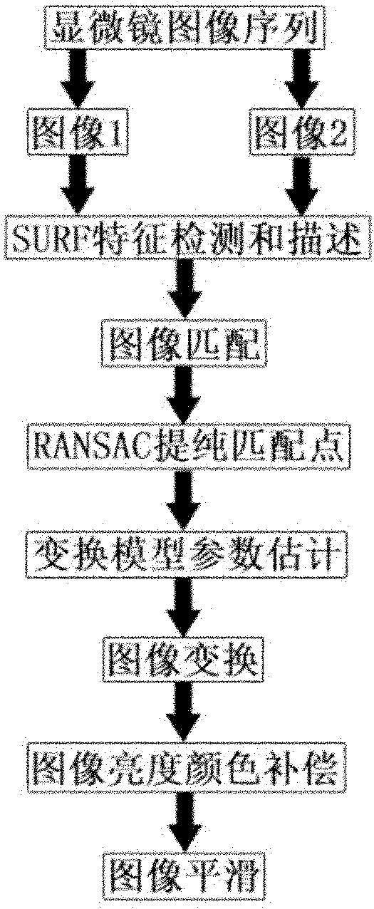
[0051] Introduce an image stitching system based on SURF operands in the field of high-magnification automatic microscope image stitching. The whole system includes the acquisition of microscope image sequences, SURF feature detection and description, image matching, removal of mismatched points, image transformation, color brightness compensation and image smoothing, etc. Step, multiple high-resolution images can be spliced ​​into a larger high-resolution panorama, so that the panorama can not only reflect the panoramic information of the microscope image, but also can well express the detailed information of the microscope image.

[0052] Such as figure 1 As shown, the implementation steps of the microscope image stitching method based on the SURF operand proposed by the present invention are as follows:

[0053] 1) Utilize the Hessian matrix to obtain the...


PUM


Abstract

The invention discloses a speeded up robust features (SURF) operand-based microscope image splicing method. In the method, an SURF operand is introduced in the field of microscope image splicing aiming at a large number of spot-like structures such as bubbles, cells and the like in a medical microscope image; the SURF operand is improved; an area is segmented and simultaneously a Gaussian weighting process is simulated; and the number of dimensions of a description vector is halved; therefore, the computational overhead for matching feature points is reduced. By applying the method, the matching accuracy is ensured and simultaneously the speed is improved greatly. The method can be suitable for the field of medical image processing and other computer vision research fields related to image splicing.

Description

technical field [0001] The invention relates to a method for mosaicing microscope images based on SURF operands. Background technique [0002] A microscope is a widely used optical instrument. It enlarges the observed object through the function of the optical system, displays the microscopic structure, and understands and studies the characteristics of the object from the microscopic form. At present, it has been widely used in various fields such as biology, pathology, cell histology, and medicinal chemistry. The field of view of the microscope is small, and images of a large field of view cannot be collected at one time. At the same time, for data measurement of large targets, incomplete images need to be spliced ​​into a whole. Therefore, splicing adjacent images together becomes a key link in microscope measurement. [0003] The image of the microscope shrinks with the increase of the magnification. Under the low magnification lens, the field of view is larger but it ...

Claims


Application Information

Patent Timeline
Patent Type & Authority Applications(China)
IPC IPC(8): G06T11/60G02B21/36
Inventor 陈辉李哲
Owner SHANDONG UNIV
Learn More