
Mask construction for cardiac subtraction

A technology of digital subtraction and mask image, which is applied in the direction of radiological diagnostic instruments, applications, cardiac catheterization, etc., can solve problems such as residual motion difficulties, and achieve the effect of reducing artifacts

Active Publication Date: 2011-07-20
KONINKLIJKE PHILIPS NV
View PDF5 Cites 8 Cited by
  • Summary
  • Abstract
  • Description
  • Claims
  • Application Information

AI Technical Summary

Problems solved by technology

As a result, it is much more difficult to compensate for residual motion in cardiac DSA compared to non-cardiac DSA
But it is critical to limit as much as possible the artifacts that spoil the subtracted image presented to the clinician

Method used


Image

  • Mask construction for cardiac subtraction
  • Mask construction for cardiac subtraction
  • Mask construction for cardiac subtraction

Examples


Embodiment Construction

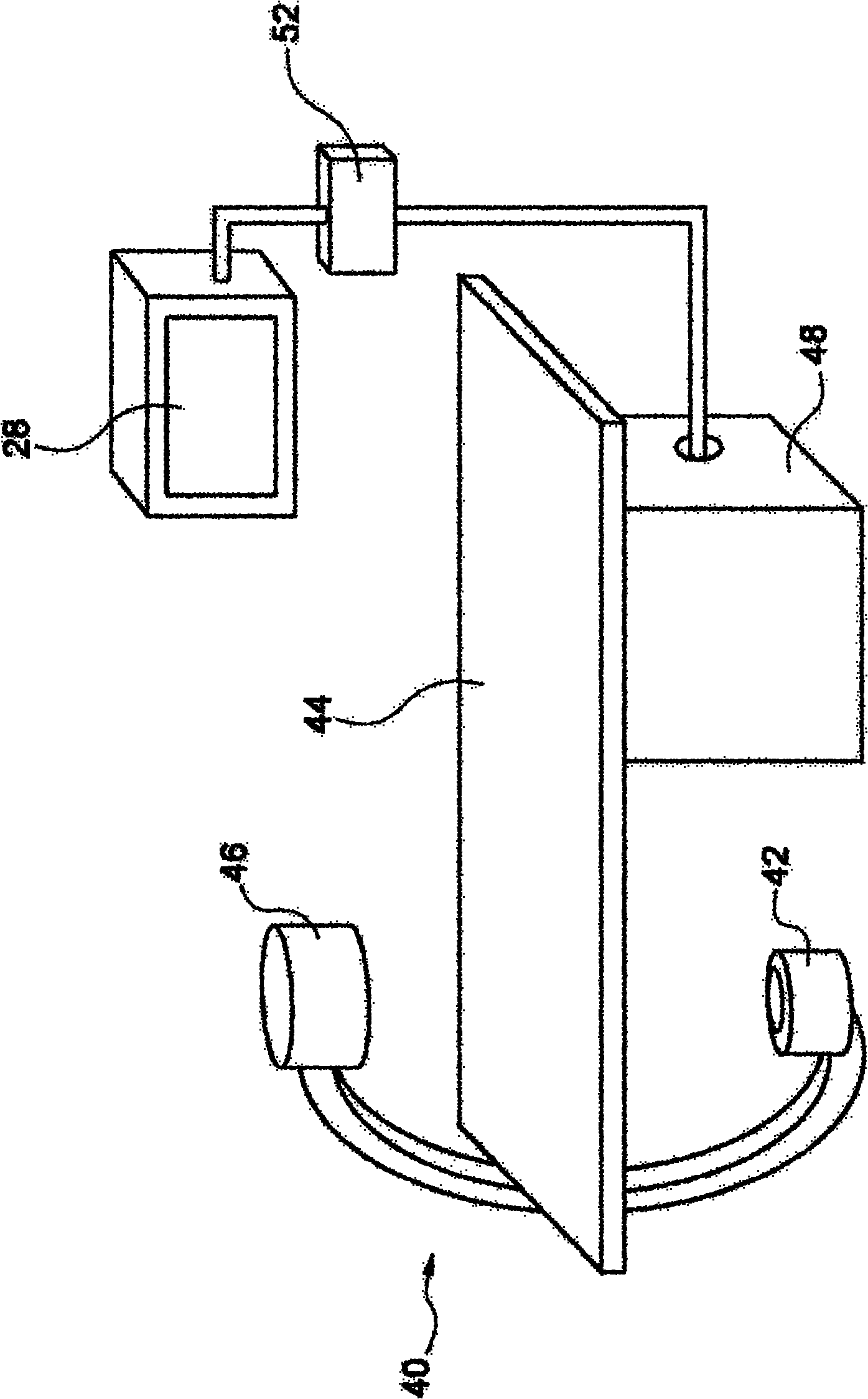
[0026] figure 1 An X-ray imaging system 40 is shown schematically. An X-ray radiation source 42 is provided to generate X-ray radiation. A station 44 is provided for receiving subjects to be examined. Furthermore, the X-ray image detection module 46 is opposite the X-ray radiation source 42 , ie the subject is located between the X-ray radiation source 42 and the detection module 46 during the radiation procedure. The latter sends data to a data processing unit 48 connected to the detection module 46 and to the radiation source 42 . Furthermore, a display 28 is arranged near the table 44 to display information to the person operating the x-ray imaging system, ie the clinician. Preferably, the display 28 is movably mounted for individual adjustment based on inspection conditions. Also, the interface unit 52 is arranged to input information by the user. Basically, the image detection module 46 generates images by exposing the subject to X-ray radiation, which are further pr...


PUM


Abstract

In order to provide an improved method for obtaining a DSA image in which a residual motion effect in cardiac Digital Subtraction Angiography (DSA) is reduced during a perfusion stage, and also to display a subtraction image containing less motion artifact, a method for performing Digital Subtraction Angiography (DSA) in an imaging device comprises the follow steps of: generating a first image sequence of a mask image (10) of a person to be checked; generating at least a first radiography image (22) in a first stage (16), in the first radiography image (22) a portion of the mask image (10) of the person to be checked having a contrast different from that of the first image sequence; subtracting the mask image (10) from at least one first radiography image (22) to generate a first DSA image sequence (24); subtracting a DSA image of the first DSA image sequence (24) from the first radiography image (22) to generate an extended mask image sequence (32); in a second stage (18), generating a second radiography image (34) using the image device, wherein the first and second stages are divided by predetermined phasing time limit (20); subtracting an image of the extended mask image sequence (32) from the second radiography image (34) to generating a second DSA image sequence (38); and displaying the second DSA image sequence (38) on a display (28).

Description

technical field [0001] The present invention relates to perfusion procedures in digital subtraction angiography (DSA), especially cardiac DSA. The invention also relates to an imaging system implementing DSA, a computer program element and a computer readable medium. Background technique [0002] For some human examinations, there is a great advantage in imaging only blood vessels. A known method for this purpose is subtraction angiography, which is based on a perfusion procedure. Basically, first and second images of the region of interest are acquired. A contrast agent is introduced into the blood vessel between the two images, and the contrast agent absorbs the X-rays. After injection of the contrast agent, the X-ray imaging device records an angiographic sequence, which shows blood vessels containing the contrast agent, highlighted in the X-ray image. To make vascular, and especially myocardial, information more accessible to clinicians and improve its visibility in ...

Claims


Application Information

Patent Timeline
Patent Type & Authority Applications(China)
IPC IPC(8): A61B6/00
CPCA61B6/481A61B6/484A61B6/504A61B6/507A61B6/5264
Inventor R·弗洛朗V·A·奥夫雷O·博纳富
Owner KONINKLIJKE PHILIPS NV
Learn More