Quantitative analysis method for three-dimensional geometric structure of heart mitral valve device

A three-dimensional geometric and quantitative analysis technology, applied in the field of quantitative analysis of three-dimensional geometric structure, can solve the problems of limited clinical diagnosis of mitral valve three-dimensional geometric structure and function, rough shape observation, loss of three-dimensional space information, etc.

Inactive Publication Date: 2011-10-05
SHANGHAI JIAO TONG UNIV
View PDF0 Cites 11 Cited by
  • Summary
  • Abstract
  • Description
  • Claims
  • Application Information

AI Technical Summary

Problems solved by technology

However, assessment of the three-dimensional geometry and function of the mitral valve is relatively limited in clinical diagnosis
First, the reference values ​​widely used in clinical practice are generally extracted from two-dimensional echocardiography; second, traditional functional indices (such as left ventricular volume and ejection fraction, etc.) do not make full use of the rich space provided by three-dimensional data information to describe the complex anatomical structure of the mitral valve device; third, although studies have shown that the saddle-shaped structure of the mitral valve annulus is closely related to its motor function, most of the actual clinical diagnosis is based on rough morphological observations; finally, from From the perspective of anatomical structure and functional integrity, the mitral valve device is composed of the mitral valve annulus, valve leaflets, papillary muscles, and chordae tendinees. Relatively comprehensive analysis of mitral valve devices
[0003] After searching the prior art, it was found that P.Nordblom and O.Bech-Hanssen wrote in "Echocardiography: a Journal Of Cardiovascular Ultrasound and Allies Technology" (Echocardiography: Cardiovascular Ultrasound and Related Technologies) (2007, 24-7: 665-672) article "Reference Values ​​Describing the Normal Mitral Valve and the Position of the Papillary Muscles" ("Reference Values ​​Describing the Normal Mitral Valve and the Position of the Papillary Muscles") uses two-dimensional ultrasound images, and the selected reference values All are measured on two-dimensional cross-sections, and three-dimensional space information is lost, so they are only suitable for quantitative analysis of mitral valve devices by two-dimensional ultrasound equipment

Method used


Image

  • Quantitative analysis method for three-dimensional geometric structure of heart mitral valve device
  • Quantitative analysis method for three-dimensional geometric structure of heart mitral valve device
  • Quantitative analysis method for three-dimensional geometric structure of heart mitral valve device

Examples


Embodiment Construction

[0027] The embodiments of the present invention are described in detail below. This embodiment is implemented on the premise of the technical solution of the present invention, and detailed implementation methods and specific operating procedures are provided, but the protection scope of the present invention is not limited to the following implementation example.

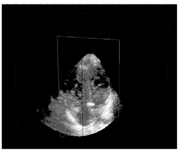
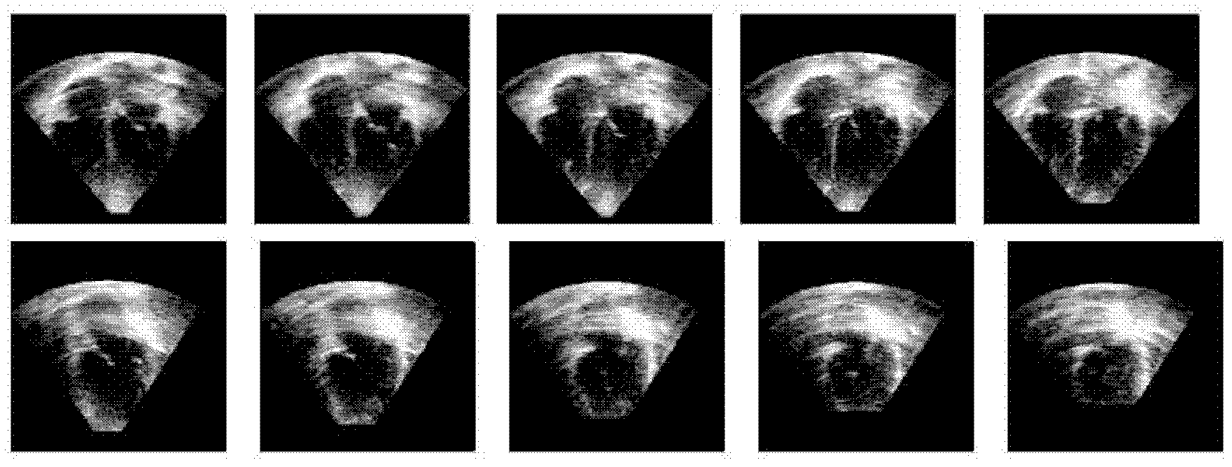
[0028] The application environment of the following example is a Philips Sonos7500 real-time three-dimensional ultrasonic diagnostic instrument and a desktop computer with IntelPentium IV 2.4GHz and 2G memory, provided that the three-dimensional matrix (matrix) probe is located at the left apex of the heart to collect Full-volume data and can clearly distinguish two The difference between the cusp apparatus and the background. For further details, take the example of full volume data collected during any whole cardiac cycle:

[0029] (1) The size of the data collected by Philips Sonos7500 real-time three-dimension...


PUM


Abstract

The invention discloses a quantitative analysis method for a three-dimensional geometric structure of a heart mitral valve device in the technical field of computer application. Quantitative analysis is realized by establishing a three-dimensional simplified model of the mitral valve device and using three-dimensional geometric structure parameters of a local coordinate system of the mitral valve device as the input of a support vector machine-based classification system. According to the method, reconstruction of the mitral valve device is not affected by an imaging mode, and the simplified three-dimensional model of the mitral valve device is obtained. The local coordinate system of the mitral valve device is established on the basis of simplified expression, so that the mitral valve device is independent of the influence factors such as the position of a sampling probe, heart motion displacement and displacement of a detected main body and the like, the three-dimensional geometric structure parameters of the mitral valve device are calculated, and the structure and the function of the mitral valve device and the spatial structure relation between the components of the mitral valve device are described.

Description

technical field [0001] The invention relates to a method in the field of computer application technology, in particular to a method for quantitatively analyzing the three-dimensional geometric structure of a heart mitral valve device. Background technique [0002] Real-time three-dimensional ultrasound system is currently the main diagnostic imaging tool for diagnosing cardiac mitral valve lesions. However, the evaluation of three-dimensional geometry and function of the mitral valve is relatively limited in clinical diagnosis. First, the reference values ​​widely used in clinical practice are generally extracted from two-dimensional echocardiography; second, traditional functional indices (such as left ventricular volume and ejection fraction, etc.) do not make full use of the rich space provided by three-dimensional data information to describe the complex anatomical structure of the mitral valve device; third, although studies have shown that the saddle-shaped structure ...

Claims


Application Information

Patent Timeline
Patent Type & Authority Applications(China)
IPC IPC(8): G06F19/00A61B8/08
Inventor 宋薇杨新孙锟王静邱俊蔚
Owner SHANGHAI JIAO TONG UNIV
Why Eureka
Try Eureka