
Capsule-type medical device

A medical device and capsule technology, applied in the field of capsule medical devices, can solve problems such as inability to inject drugs, and achieve the effect of reliable injection

Inactive Publication Date: 2013-12-18
OLYMPUS CORP
View PDF7 Cites 0 Cited by
  • Summary
  • Abstract
  • Description
  • Claims
  • Application Information

AI Technical Summary

Problems solved by technology

[0007] However, depending on the puncture state of the injection needle that punctures the living tissue, there is a problem that it may not be possible to reliably inject the drug into the living tissue.

Method used


Image

  • Capsule-type medical device
  • Capsule-type medical device
  • Capsule-type medical device

Examples


Embodiment approach 1

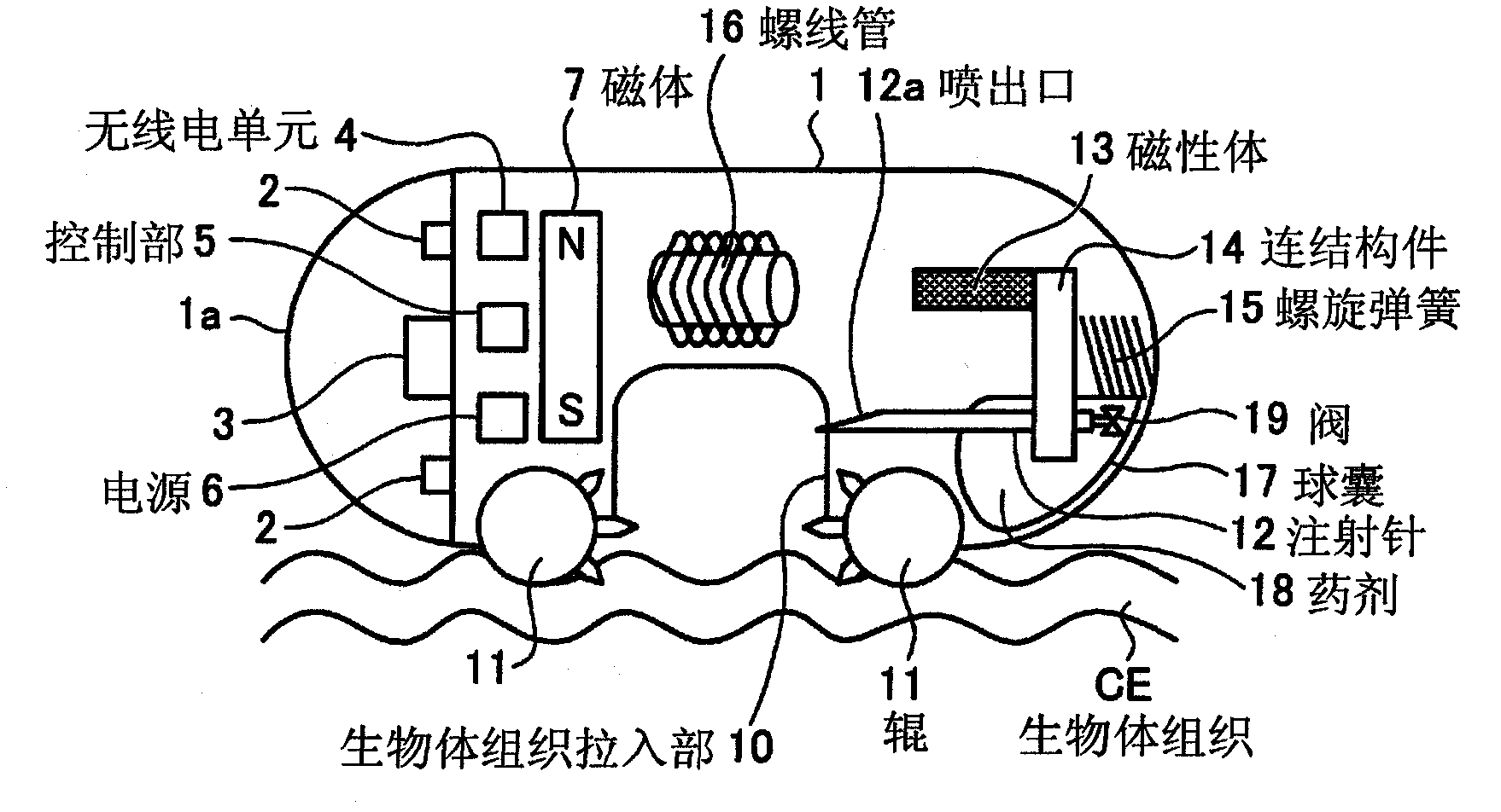
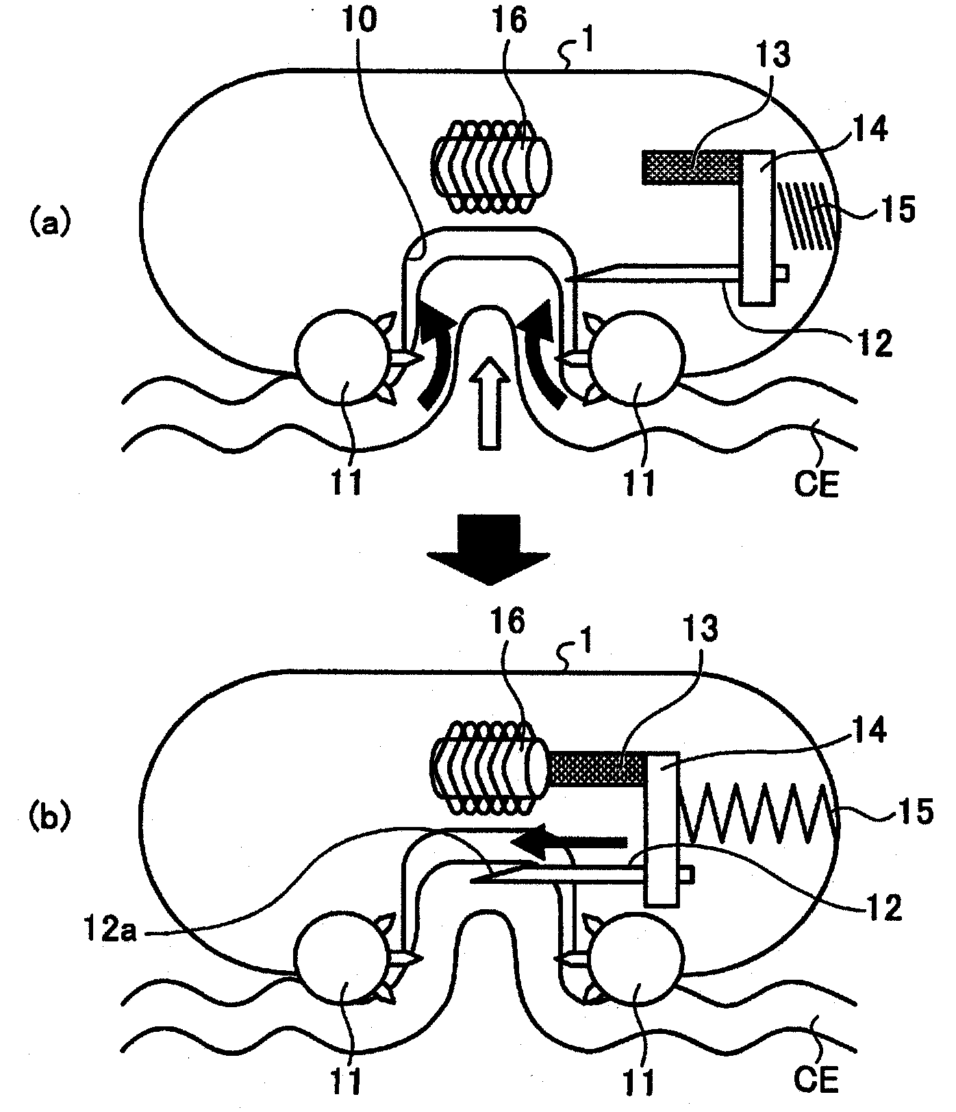
[0039] figure 1 It is a schematic diagram showing the configuration of the capsule medical device according to Embodiment 1 of the present invention. Such as figure 1 As shown, the capsule medical device is a capsule-shaped medical device of a size that can be introduced into the subject. It is introduced into the subject, and the target biological tissue is drawn into the capsule medical device. The medicine is injected into the pulled living tissue, and then the drawing of the living tissue injected with the medicine is released.

[0040] In this capsule medical device, one end opening of the cylindrical case is closed by a transparent dome-shaped case 1a, and various functions are incorporated in the capsule-shaped case 1 maintained in a liquid-tight state. Near the dome-shaped casing 1a, there is an imaging unit 3 on the axis of the capsule-shaped casing 1. The imaging unit 3 is realized by an imaging element, a condensing optical system, etc., and captures in-vivo ima...

Embodiment approach 2

[0056] Next, Embodiment 2 of the present invention will be described. Such as Figure 5 As shown, in Embodiment 2, the living tissue drawing-in portion 30 corresponding to the living tissue drawing-in portion 10 is formed in the inside of the capsule-shaped housing 1 in a partially opened cylindrical shape. Furthermore, the roll-in part 33 is attached to the tip portion of the arm 31, and the injection needle 34 is attached to the tip portion of the arm 32, the arm 31 rotates around the axis C1 of the cylinder, and the arm 32 rotates around the axis C1, It is substantially perpendicular to the arm 31 and rotates 90 degrees later than the arm 31 in the rotation direction. In addition, the piercing direction of the injection needle 34 faces the circumferential direction of the rotation direction. That is, the injection needle 34 faces the tangential direction of the rotation locus of the tip of the arm 32 . In addition, the ejection port of the injection needle 34 is provided...

Embodiment approach 3

[0063] Next, Embodiment 3 of the present invention will be described. In both of the first and second embodiments described above, the living tissue CE is pulled in by the rotation mechanism, but in this third embodiment, the living tissue CE is pulled in by the suction mechanism.

[0064] Figure 9 It is a schematic diagram showing the schematic configuration of the capsule medical device according to the third embodiment. This capsule medical device has a living tissue suction part 40 corresponding to the living tissue suction part 10 , and a mucosal suction port 41 for sucking the living tissue CE is formed in the capsule casing 1 . In addition, the capsule housing 1 is provided with: a decompression device 43 for drawing the living tissue CE into the living tissue drawing part 40 by suction; an injection needle driving part 50 for protruding the injection needle 51 to the living body A predetermined position in the body tissue drawing-in part 40; a cam device 44, which c...


PUM


Abstract

The present invention provides a capsule-type medical device, which is introduced into a subject to inject medicine into the biological tissue in the subject. The capsule-type medical device includes: A space for pulling in the living tissue (CE) is formed in the housing (1); a roller (11) moves the living body tissue (CE) to the living body tissue drawing part (10); an injection needle (12) Protrude so that the ejection port (12a) of the medicine is located in the biological tissue drawing part (10); a magnetic body (13) and a connecting member for driving the injection needle (12) and protruding the injection needle (12) (14), coil spring (15), and solenoid (16).

Description

technical field [0001] The present invention relates to a capsule-type medical device capable of being introduced into a subject and injecting a drug into living tissue in the subject. Background technique [0002] In recent years, in the field of endoscopes, a capsule-type subject-introducing device (such as a capsule-type endoscope) equipped with an imaging function and a wireless communication function has been proposed. An in-subject introduction system for acquiring images inside a subject. In order to observe (inspect) the inside of the subject, the capsule endoscope is swallowed, for example, through the subject's mouth, and then moves around in the body cavity such as the stomach, the small intestine, and other organs until it is naturally excreted. moves while moving, and takes images inside the subject at intervals of, for example, 0.5 seconds. [0003] While the capsule endoscope is moving within the subject, an external image display device receives images capt...

Claims


Application Information

Patent Timeline
Patent Type & Authority Patents(China)
IPC IPC(8): A61B1/00A61B5/07
CPCA61B1/00082A61B1/00087A61B1/041A61M5/14248A61M5/148A61M31/002A61M2005/14252A61B34/73
Inventor 田中慎介河野宏尚泷泽宽伸片山美穗铃木启太
Owner OLYMPUS CORP
Learn More