Method for separating and culturing pig spermatogonial stem cells by mechanical process

A technique for separating and culturing spermatogonial stem cells, applied in the field of mechanical separation and culturing of porcine spermatogonial stem cells, achieving the effects of high success rate, large number of cells, and reduced experimental costs

Inactive Publication Date: 2012-01-11
SOUTH CHINA AGRI UNIV
View PDF0 Cites 3 Cited by
  • Summary
  • Abstract
  • Description
  • Claims
  • Application Information

AI Technical Summary

Problems solved by technology

However, there are deficiencies in each method

Method used


Image

  • Method for separating and culturing pig spermatogonial stem cells by mechanical process
  • Method for separating and culturing pig spermatogonial stem cells by mechanical process
  • Method for separating and culturing pig spermatogonial stem cells by mechanical process

Examples


Embodiment 1

[0030] Example 1 Acquisition and cultivation of porcine spermatogonia

[0031] (1) On the ultra-clean bench, put the piglet’s testes into a petri dish filled with PBS, and wash with PBS for 2 to 3 times to remove blood stains;

[0032] (2) Remove the testicular tunica, remove the connective tissue in the testicular parenchyma with sharp forceps, and cut the remaining testicular tissue into pieces with iris scissors until the tiny tissue pieces are no longer visible, then transfer to a 15 ml centrifuge tube;

[0033] (3) Add 10 times the volume of PBS, pipette gently, then centrifuge at 16°C, 600 rpm for 5 min, and discard the supernatant. Repeat this process for 2-3 times;

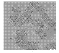
[0034] (4) Adjust the number of seminiferous tubules and inoculate into 25 cm pre-coated with 0.1% gelatin 2 Culture flasks at 37 °C, 5 % CO 2 , saturated humidity sterile incubator conditions ( Figure 1~5 ).

Embodiment 2

[0035] Example 2 Positive detection of alkaline phosphatase (Alkaline Phosphatase, AP)

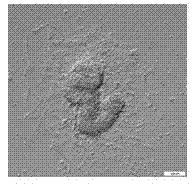
[0036] Wash the PSSCs to be detected once with PBS; fix with 4% paraformaldehyde at room temperature for 20 min, wash with PBS three times, each time for 5 min; add 1 mL of alkaline phosphatase buffer, and add 6.6 μL of NBT and BCIP (vector) 3.3 μl each, make a mixing tube, mix thoroughly; incubate at room temperature with light blocked for 10-15 min, wash with PBS, stop the reaction in time, observe and take pictures under a microscope (such as Figure 6 ).

[0037] Through AP staining, it can be preliminarily judged that the clones obtained by the one-step enzymatic method are PSSCs clones.

Embodiment 3

[0038] Example 3 Immunofluorescence identification and detection of PSSCs

[0039] (1) After the cell slides are prepared, take out the slides and wash them with PBS at 37 ℃ for 3 times, each time for 3-5 s;

[0040] (2) Fix with 4% paraformaldehyde for 10-30 min; wash with PBS twice, 5 min each time;

[0041] (3) 0.5 % Triton perforated and perforated membrane for 15 minutes; rinsed with PBS twice, 5 minutes each time;

[0042] (4) Block with 1% BSA for 30 min (do not wash after blocking);

[0043] (5) Add primary antibody diluted with 1% BSA, overnight at 4°C; wash with PBS twice, 5 min each time;

[0044] (6) Add secondary antibody diluted with 1 % BSA, at 37 ℃ / 1 h; wash with PBS twice, 5 min each time;

[0045] (7) Stain with 10 μg / mL Hochest for 5 min; mount the slides with anti-fade, and take pictures.

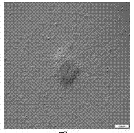
[0046] The clonal group obtained by immunofluorescence detection mechanism specifically expresses the important stem cell marker gene Oct4, which proves that it is...


PUM


Abstract

The invention discloses a method for separating and culturing pig spermatogonial stem cells by a mechanical process. The method of the invention is characterized in that the characteristics of animal testicular tissue are combined, and the testicular tissue can be separated by the mechanical process and then cultured, so that the method for separating and purifying spermatogonial stem cells can be enriched. According to the invention, the pig spermatogonial stem cells can be obtained without enzyme digestion, the method of the invention has the advantages of fast cells clone and good state ofcells, and solves the problems of difficult separation, small quantity and high requirement of culture condition of spermatogonial stem cells. Compared with the separating technology of the prior art, the method of the invention has simple operation, and is capable of reducing the difficulty to obtain cells, substantially reducing the experimental cost and raising the success rate of the experiments.

Description

technical field [0001] The invention relates to the field of cell separation and culture, in particular to a method for mechanically separating and culturing porcine spermatogonial stem cells. Background technique [0002] Spermatogonial stem cells (SSCs) are a type of undifferentiated type A spermatogonia located on the basement membrane of the seminiferous tubules of male animals, which can not only self-renew and maintain a constant number of their own population, but also be able to differentiate and finally produce sperm cells, and the only replicable multipotent diploid cells in adults. [0003] The methods of isolating SSCs in vitro mainly focus on the combination of mechanical method and enzymatic digestion method. Now SSCs are mainly obtained by enzymatic digestion, and then single SSCs are finally obtained through various purification methods. Whether the purified SSCs are more conducive to the proliferation of SSCs is also There are different opinions. At presen...

Claims


Application Information

Patent Timeline
Patent Type & Authority Applications(China)
IPC IPC(8): C12N5/076
Inventor 张守全白银山
Owner SOUTH CHINA AGRI UNIV
Why Eureka
Try Eureka