
Method and device for removing scanning table from CTA (Computed Tomography Angiography) image

A technology of image center and scanning bed, which is applied in the field of computer, can solve the problems affecting the observation of blood vessels, etc., and achieve the effect of accurate observation, short time and fast operation speed

Inactive Publication Date: 2012-01-18
NEUSOFT CORP
View PDF2 Cites 9 Cited by
  • Summary
  • Abstract
  • Description
  • Claims
  • Application Information

AI Technical Summary

Problems solved by technology

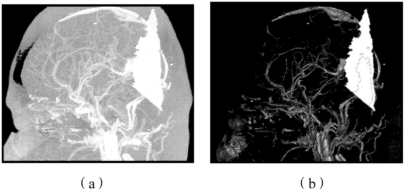
from figure 1 It can be seen from the figure that part of the scanning bed image still remains after the subtraction operation of the CTA image, so that a high-brightness bed image will appear in the 3D visualization reconstruction results, which will affect the observation of blood vessels. ignored this question

Method used


Image

  • Method and device for removing scanning table from CTA (Computed Tomography Angiography) image
  • Method and device for removing scanning table from CTA (Computed Tomography Angiography) image
  • Method and device for removing scanning table from CTA (Computed Tomography Angiography) image

Examples


Embodiment Construction

[0056]The following will clearly and completely describe the technical solutions in the embodiments of the present invention with reference to the accompanying drawings in the embodiments of the present invention. Obviously, the described embodiments are only some, not all, embodiments of the present invention. Based on the embodiments of the present invention, all other embodiments obtained by persons of ordinary skill in the art without creative efforts fall within the protection scope of the present invention.

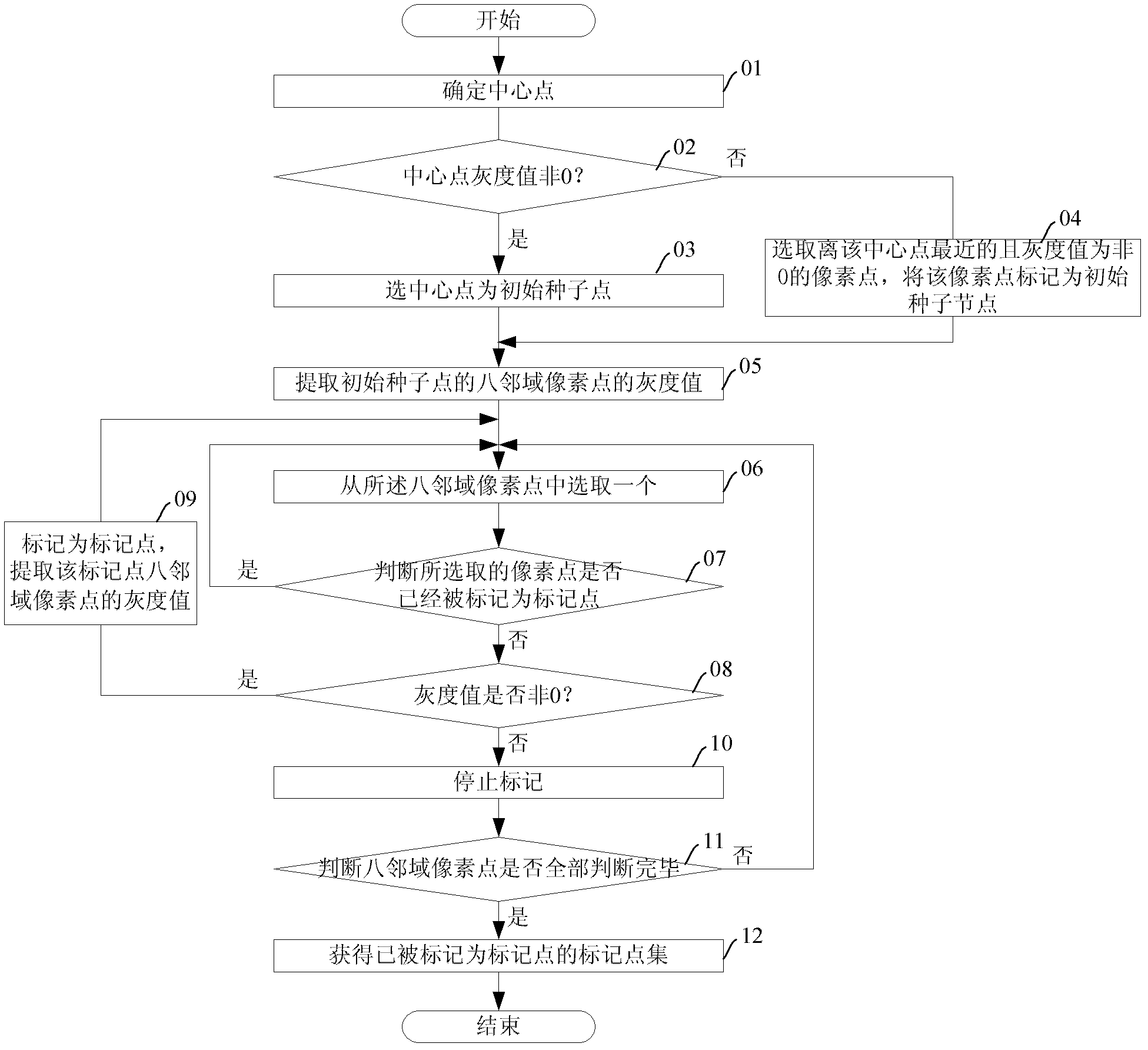
[0057] The present invention considers that the gray value range presented by the scanning bed body in the tomographic image partially overlaps with the gray value range of the brain bone, so it cannot be removed by threshold segmentation. However, since there is a certain air gap between the scanning bed image and the brain tissue image, the bed image is not connected to the brain tissue area, so the present invention uses the method of region growth to extract the ...


PUM


Abstract

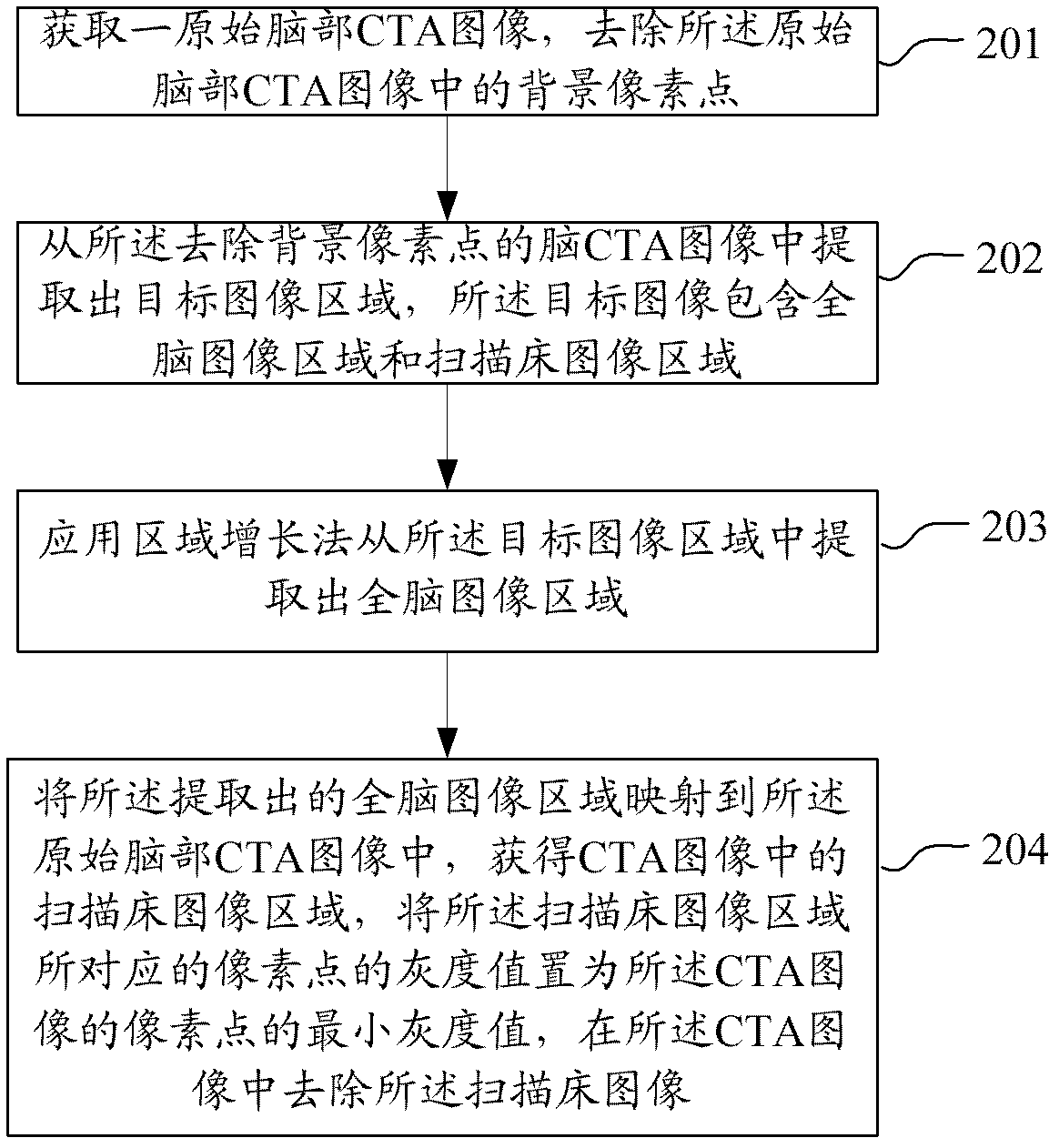
The application discloses a method and device for removing a scanning table from a CTA (Computed Tomography Angiography) image. The method comprises the following steps of: acquiring a brain CTA image, and removing background pixel points in the CTA image; extracting a whole-brain image from the brain CTA image with the background pixel points being removed; finding a pixel point corresponding to a scanning table image from the whole-brain image by using a region growing algorithm, setting the gray value of the pixel point corresponding to the scanning table image to be zero, and removing the scanning table image. By using the method and device for removing the scanning table from the CTA image, the scanning table image can be completely removed, therefore, the problem that partial scanning table image is still remained after the CTA image is subjected to subtraction operation is avoided, in addition, the problem of a table image with high brightness in a three-dimensional visualized reconstruction result is avoided, clear and foreign-matter-free brain image data is provided for three-dimensional reconstruction of brain vessels, and more accuracy and efficiency in observing the vessels is realized. In addition, the arithmetic speed is high and the time is short.

Description

technical field [0001] The invention relates to the field of computer technology, in particular to a method and device for removing a scanning bed from a CTA image. Background technique [0002] CT is a full-featured disease detection instrument, which is the abbreviation of computerized X-ray tomography. CT images are represented by different gray scales. In order to display the whole organ, multiple continuous slice images are required. [0003] Currently, in clinical practice, multi-slice spiral CT angiography (MSCTA, multi-slice spiral CT angiography) is a new type of angiography technique, and the images obtained by this technique are called CTA images. The technology is non-invasive to the patient and provides a convenient and reliable basis for doctors to observe and diagnose cerebrovascular diseases. [0004] Intracranial vascular images are usually obtained by registration and subtraction of CT images and CTA images. The principle is to register the brain CT imag...

Claims


Application Information

Patent Timeline
Patent Type & Authority Applications(China)
IPC IPC(8): G06T5/00
Inventor 杨金柱韩芳芳覃文军赵大哲栗伟袁玉亮任福龙
Owner NEUSOFT CORP
Learn More