Eye fundus image structure segmentation method based on full convolution neural network

A technology of convolutional neural network and fundus image, applied in the field of structure segmentation of fundus image based on full convolutional neural network, to achieve the effect of improving accuracy and rapid observation

Active Publication Date: 2019-02-12
UNIV OF ELECTRONICS SCI & TECH OF CHINA
View PDF5 Cites 21 Cited by
  • Summary
  • Abstract
  • Description
  • Claims
  • Application Information

AI Technical Summary

Problems solved by technology

However, how to apply fully convolutional neural network to fundus image segmentation needs further research.

Method used


Image

  • Eye fundus image structure segmentation method based on full convolution neural network
  • Eye fundus image structure segmentation method based on full convolution neural network
  • Eye fundus image structure segmentation method based on full convolution neural network

Examples


Embodiment

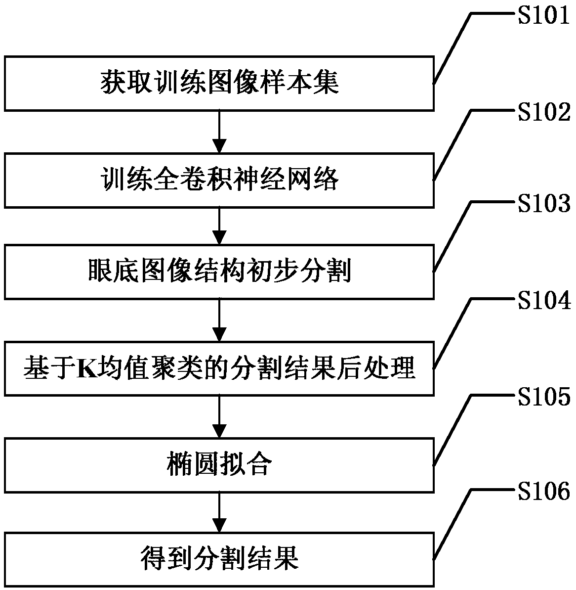
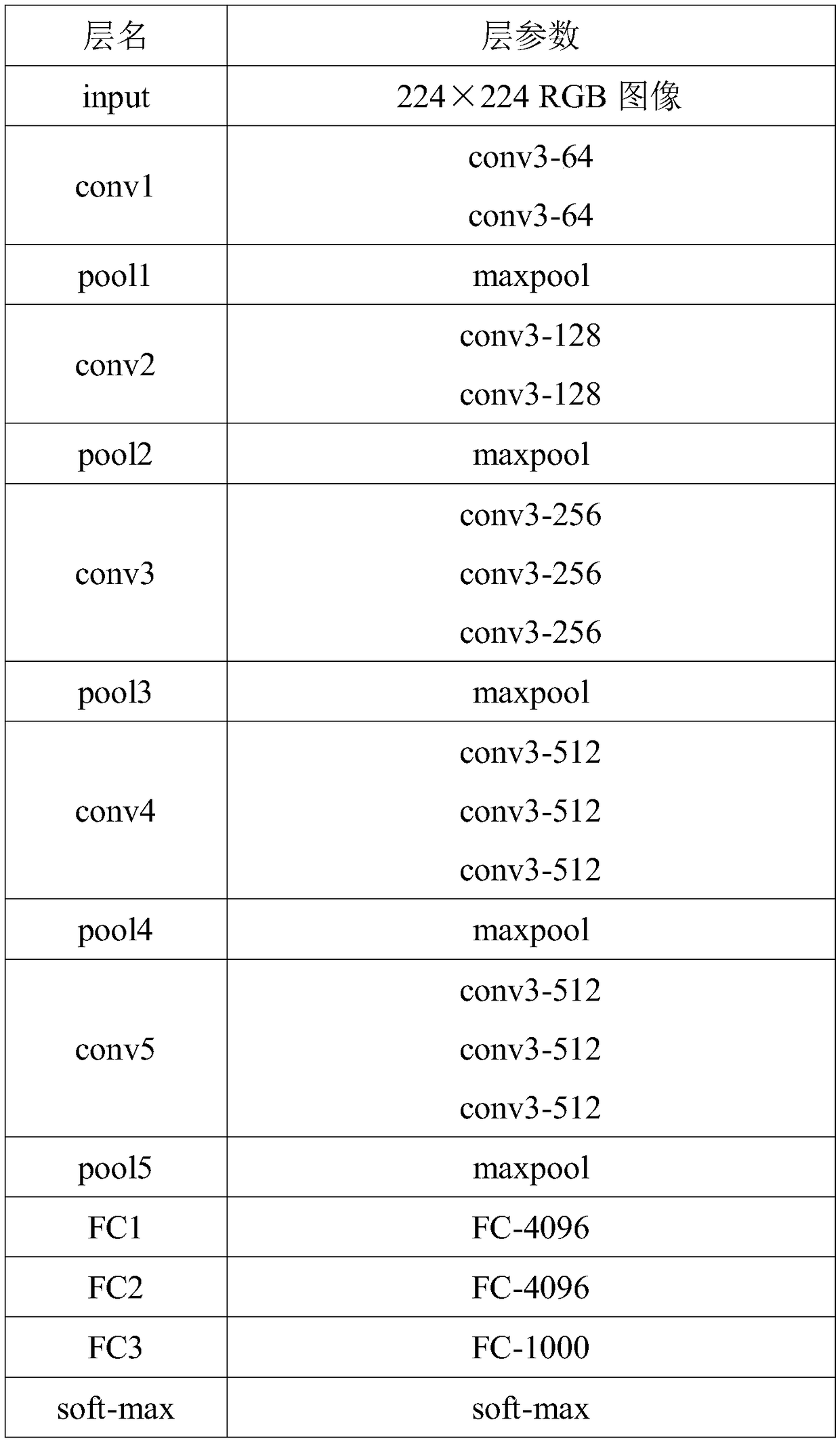
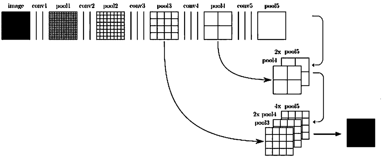
[0024] figure 1It is a flow chart of a specific embodiment of the method for segmenting fundus image structure based on a fully convolutional neural network in the present invention. Such as figure 1 As shown, the specific steps of the fundus image structure segmentation method based on the full convolutional neural network of the present invention include:

[0025] S101: Obtain a training image sample set:

[0026] Obtain several fundus image samples, normalize each fundus image sample to a preset size, mark the three target structures of the macula, optic disc, and optic cup in each fundus image sample, and generate according to the corresponding target structure labeling information The target result map, except the pixels belonging to the target structure are the original pixels in the target result map, all other pixels are set as background pixels. Each fundus image sample and the corresponding target result map are regarded as a pair of training image samples, so as ...


PUM


Abstract

The invention discloses an eye fundus image structure segmentation method based on full convolution neural network. The method comprises the steps of firstly marking the macula, optic disc and optic cup of each fundus image sample, and generating the corresponding target result map of each fundus image sample; then using each fundus image sample and corresponding target result map as a pair of training image samples to obtain the training image sample set; using the training image sample set to train the full convolution neural network, and inputting the fundus image to be segmented to the full convolution neural network to get the corresponding target result map; then using the K-means clustering and ellipse fitting to get the segmentation result. The method of the invention can improve the precision of the fundus image segmentation and better distinguish the normal physiological structure of the fundus.

Description

technical field [0001] The invention belongs to the technical field of fundus image processing, and more specifically relates to a fundus image structure segmentation method based on a fully convolutional neural network. Background technique [0002] Fundus images can be used to diagnose fundus diseases such as glaucoma and fundus macular degeneration, and can also provide a reference for diagnosing diseases such as diabetes and hypertension. The traditional retinal image processing method is mainly staged, subject to the doctor's experience, time-consuming, labor-intensive, and inefficient. The parameters in the patient's fundus image can be obtained through computer-aided automatic detection, which can not only provide convenience for doctors when diagnosing diseases, but also greatly shorten the time to achieve the effect of efficient medical detection of diseases. [0003] Many scholars at home and abroad have conducted research in this area, and proposed a variety of m...

Claims


Application Information

Patent Timeline
Patent Type & Authority Applications(China)
IPC IPC(8): G06T7/00G06T7/11G06T7/187G06T7/194
CPCG06T7/0012G06T2207/20081G06T2207/20084G06T2207/30041G06T7/11G06T7/187G06T7/194
Inventor 秦臻王亚敏丁熠秦志光
Owner UNIV OF ELECTRONICS SCI & TECH OF CHINA
Why Eureka
Try Eureka