X-ray computer tomography system and method

An X-ray tube and computing module technology, applied in the field of medical imaging, can solve the problems of not changing the opening of the collimator, not adjusting the exposure area of ​​the object to be inspected, and not considering the size change and shape change of the ROI, so as to reduce the X-ray dose , reduce extra exposure, easy to achieve effect

Active Publication Date: 2012-04-04
SIEMENS SHANGHAI MEDICAL EQUIP LTD
View PDF6 Cites 12 Cited by
  • Summary
  • Abstract
  • Description
  • Claims
  • Application Information

AI Technical Summary

Problems solved by technology

However, this method only proposes a solution for a single rotation scan with a fixed FOM, without considering the size and shape changes of the ROI in the scanning direction, where the scanning direction is the horizontal direction of the examination bed in and out of the rack in the CT system, which is different from the The phi plane is vertical, usually in the z-axis direction
[0005] The patent application whose publication number is CN 1446517A discloses an X-ray data acquisition system, which adopts an elliptical cross section or a circular cross section, and calculates the transmission value according to the target value of the standard deviation of the image, the expected value of the standard deviation of the image and the thickness factor. The dose value of X-rays within the predetermined angle range of the body axis direction of the object to be inspected is achieved by changing the tube current of the X-ray tube, but the opening of the collimator is not changed, nor is it scanned according to the object to be inspected The size of the direction changes to adjust the exposure area of ​​the object to be inspected accordingly

Method used


Image

  • X-ray computer tomography system and method
  • X-ray computer tomography system and method
  • X-ray computer tomography system and method

Examples


Embodiment Construction

[0035] In order to make the purpose, technical solution and advantages of the present invention clearer, the following examples are given to further describe the present invention in detail.

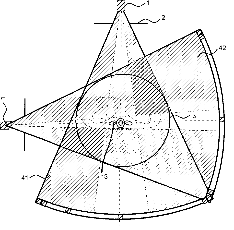
[0036] Since the cross-sections of most organs or tissues of the human body have approximately elliptical outer contours, the cross-section of the object to be inspected in the present invention is an elliptical cross-section. By calculating the major axes and The projection value of the minor axis at the detector can be deduced from the properties of similar triangles to deduce the respective major and minor axes of the plurality of elliptical sections, which also determines the flattening degree and size of each elliptical section.

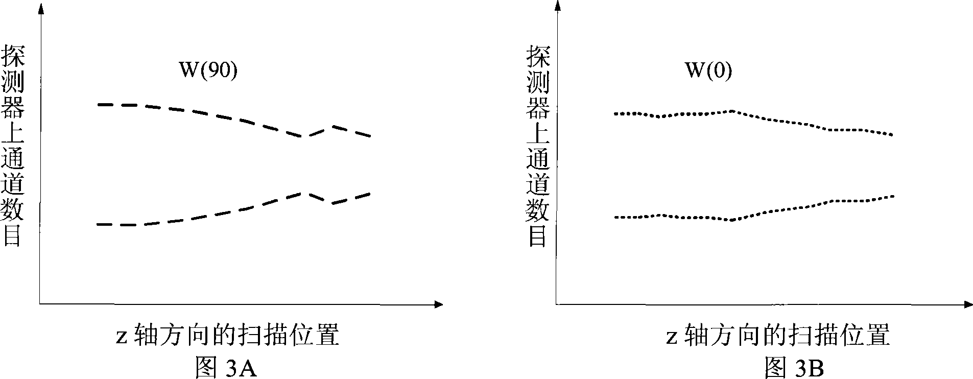
[0037] The present invention calculates the opening width of the collimator according to the different sizes of multiple elliptical sections in the scanning direction of the object to be inspected and the chord projection values ​​of the same elliptical sec...


PUM


Abstract

The invention discloses an X-ray computer tomography system, which comprises an X-ray tube, a collimator and a detector and is used for photographing a plurality of elliptical cross sections of an object to be detected in the scanning direction; the detector comprises a plurality of detector channels; the X-ray tube, the collimator and the detector synchronously rotate around the object to be detected; the collimator is provided with an adjustable opening; the X-ray computer tomography system also comprises a computation assembly and an adjustment assembly, wherein the computation assembly is used for computing opening widths of the collimator, which are required for irradiating each elliptical cross section, under different projection angles, and transmitting the opening widths to the adjustment assembly; and the adjustment assembly is used for adjusting the opening of the collimator according to the opening widths. The invention also discloses an X-ray computer tomography method. Due to adoption of the system and method disclosed by the invention, extra exposure of a surrounding area of the object to be detected can be reduced, and the X-ray dose accepted by a patient can be reduced.

Description

technical field [0001] The invention relates to the field of medical imaging, in particular to an X-ray computed tomography system and method. Background technique [0002] In X-ray computed tomography (CT) system, dose characteristic is a very important performance index. Keeping the scanning dose as low as possible has become a general rule in the development of current CT systems. In a CT system, the field of measurement (FOM) is defined as the cross-sectional area where the object to be inspected gets full radiation when the X-ray tube rotates 360 degrees in a single rotation scan, which determines the direct detection of the object to be inspected. The scan dose applied and the size of the X-ray irradiated area. [0003] Since the cross-sections of most organs or tissues of the human body have approximately elliptical outer contours, for a single rotation scan, when the X-ray tube irradiates the object to be inspected at different projection angles, the fixed circular...

Claims


Application Information

Patent Timeline
Patent Type & Authority Applications(China)
IPC IPC(8): A61B6/03
Inventor 田毅陈马昊
Owner SIEMENS SHANGHAI MEDICAL EQUIP LTD
Why Eureka
Try Eureka