

Computed tomography (CT) image and magnetic resonance (MR) image fusion method
A CT image and fusion method technology, applied in the field of fusion of CT images and MR images, can solve problems such as unsatisfactory effects, ignoring relative information, time-consuming, etc., and achieve the goal of maintaining edge and detail information, simple implementation process, and avoiding contrast Falling effect
- Summary
- Abstract
- Description
- Claims
- Application Information
AI Technical Summary
Problems solved by technology
Method used
Image

Examples
Embodiment Construction
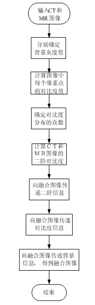
[0034] Refer to attached figure 1 , the fusion method of CT image and MR image described in the present invention comprises the following steps:
[0035] Step 1, determine the background gray value of the image:
[0036] 1.1) Statistically calculate the gray distribution of the input CT and MR images, obtain the gray histogram, and find out the gray value corresponding to the maximum peak point in the gray histogram;
[0037] 1.2) Determine whether the gray value is equal to 0, if not, use the gray value of the peak point as the background gray value of the image; if the gray value is equal to 0, use the background value of another image as the background gray value;
[0038] Step 2, according to the background gray value, use the following formula to calculate the contrast value of each pixel in the CT and MR images respectively:
[0039]
[0040] Step 3, determine the mode of each image contrast value;
[0041] 3.1) Map C(x,y) to the interval [0,255] ac...
PUM
 Abstract
Description
Claims
Application Information