Minimally invasive skeletal external fixation device for correcting foot accelerator drop and talipes varus

A technique of inversion and external fixation of the foot, applied in medical science, surgery, etc., can solve the problem of patients losing labor ability, heavy mental blow, tibial osteomyelitis, bone defect and so on

Inactive Publication Date: 2012-05-02
汤福刚
View PDF4 Cites 6 Cited by
  • Summary
  • Abstract
  • Description
  • Claims
  • Application Information

AI Technical Summary

Problems solved by technology

Seriously affect the quality of life of the patient, make it difficult for the patient to move, and even make the patient lose a certain ability to work
[0003] The cause of foot drop, foot inversion, and foot valgus is mostly due to the fact that the affected foot is in one posture for a long time during the treatment process, and the recovery is not ideal, which makes some tendons around the foot joints, such as the Achilles tendon, shorten; the scars are postoperative A common phenomenon is that some scars are stiff and atrophied; some patients recover from tibial surgery and the length of the other leg is shorter than the other leg, resulting in lameness of the patient. What is more serious is that the patient's bone is not connected after the operation or even infected due to improper handling, resulting in tibial fractures. Osteomyelitis, bone defect
[0004] At present, the Achilles tendon is mostly released for the treatment of

Method used


Image

  • Minimally invasive skeletal external fixation device for correcting foot accelerator drop and talipes varus
  • Minimally invasive skeletal external fixation device for correcting foot accelerator drop and talipes varus
  • Minimally invasive skeletal external fixation device for correcting foot accelerator drop and talipes varus

Examples


Embodiment Construction

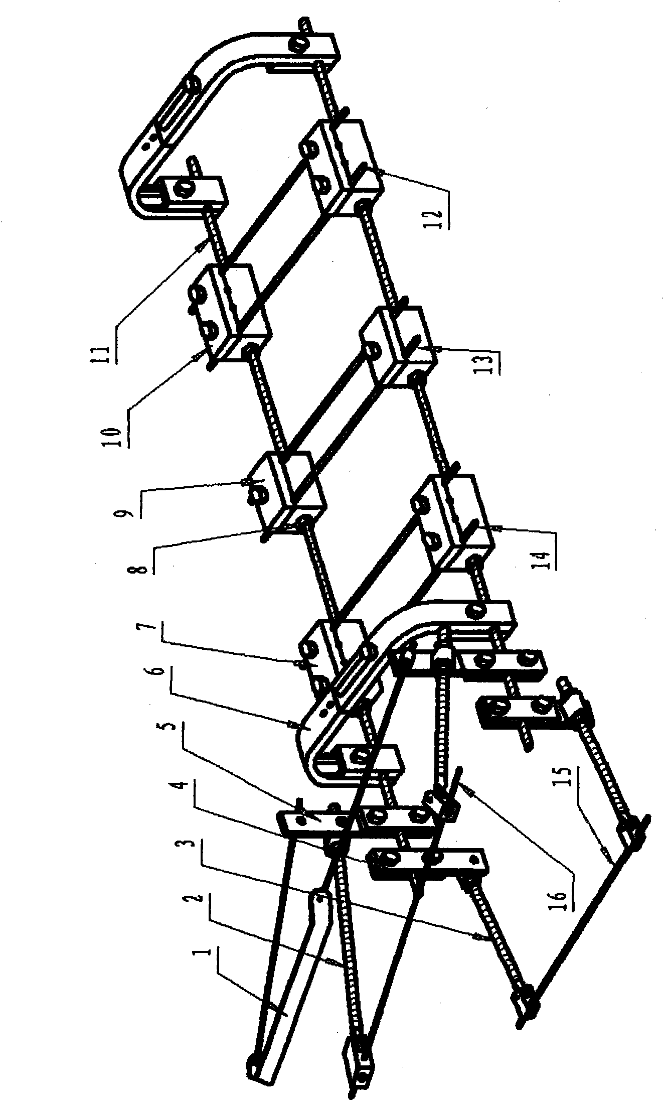
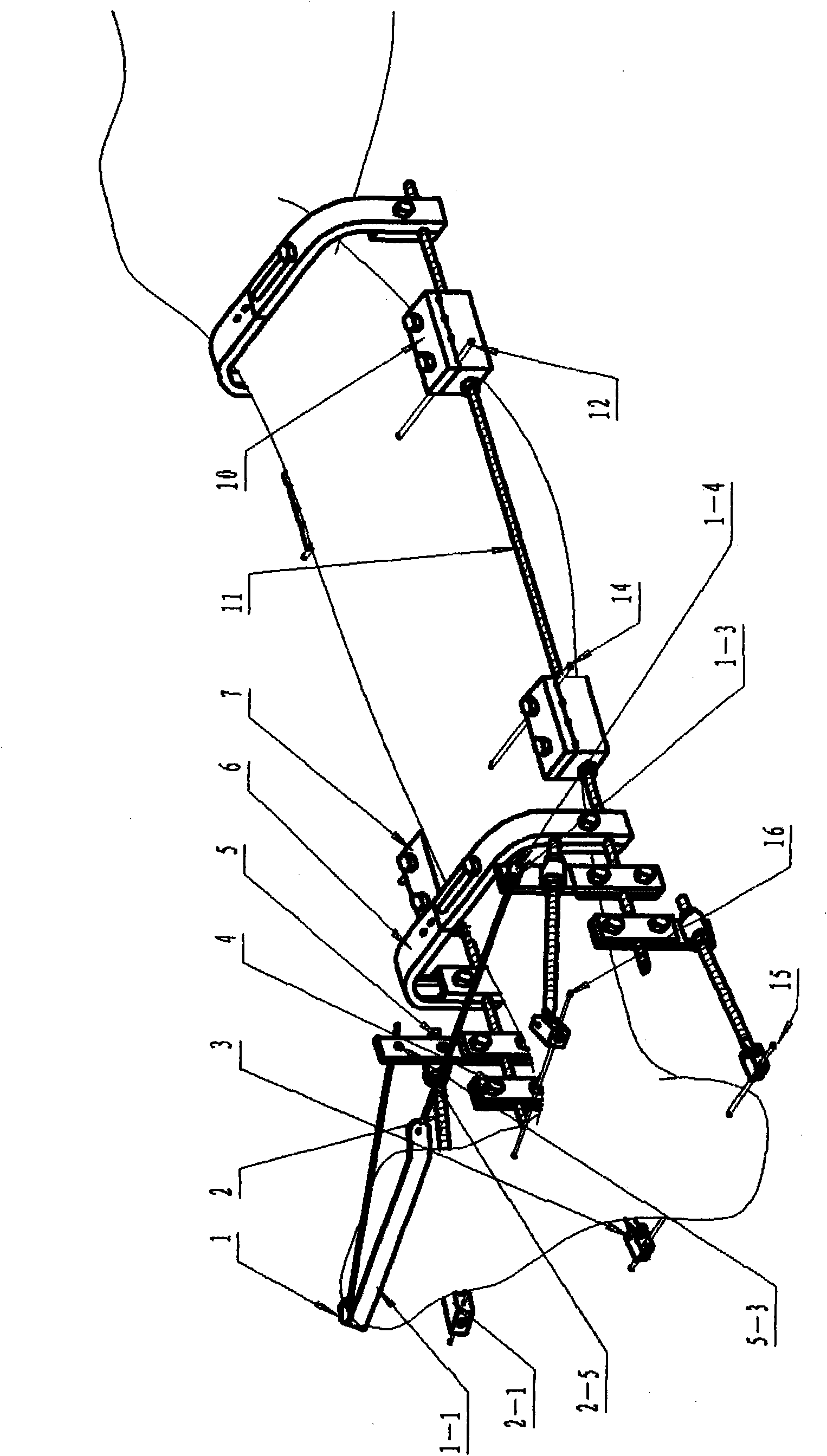
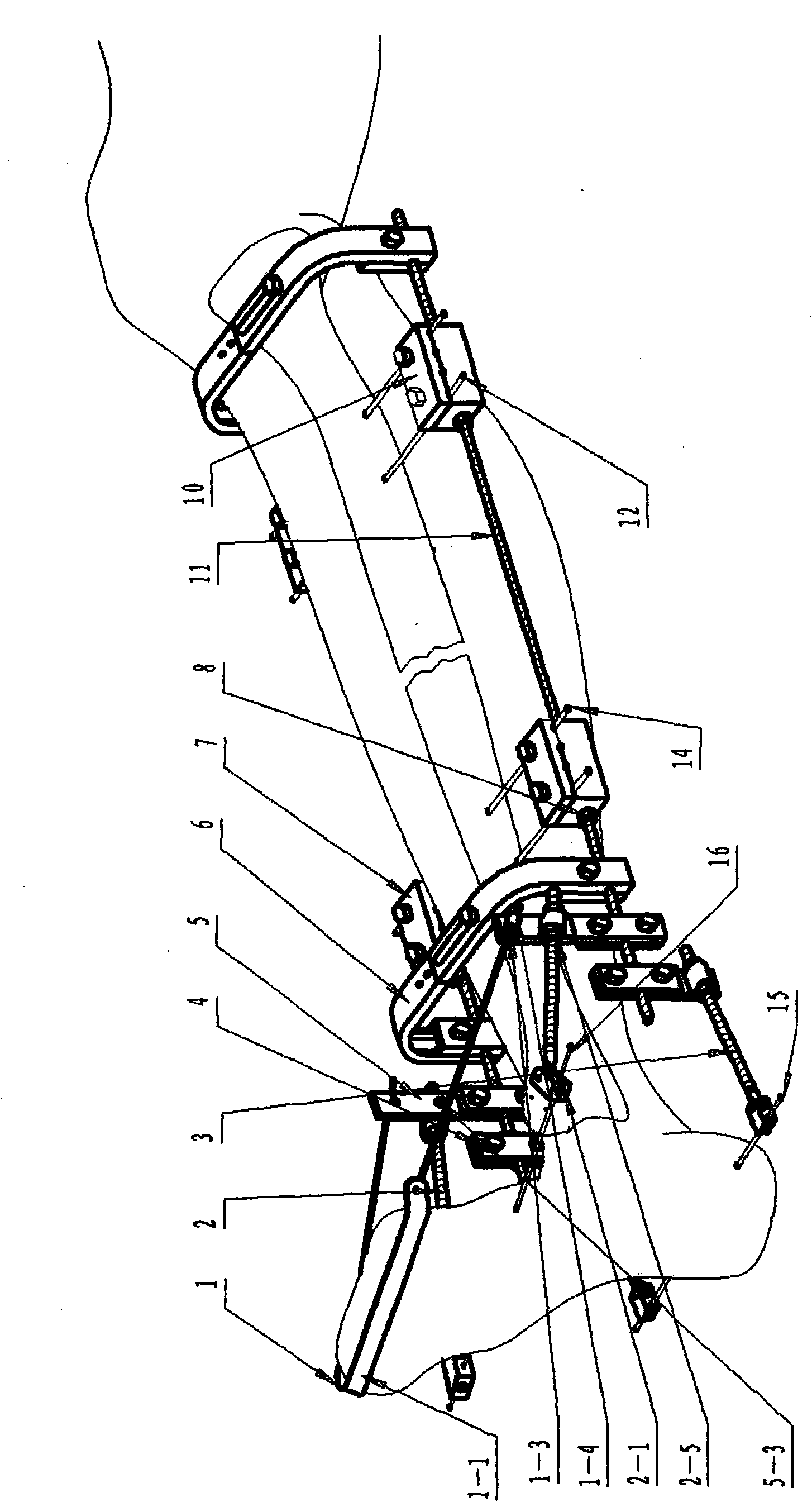
[0059] Such as figure 1 The shown minimally invasive bone external fixation device for vertical foot and foot varus correction consists of an anti-toe bending frame (1), a bone bone spicule regulator (2), a calcaneal spicule regulator (3), and a calcaneal spicule Adjuster bracket (4), Bone spicule regulator bracket (5), Bow frame (6), Distal bone spicule clip (7), Adjusting nut (8), Moving bone spicule clip (9), Proximal Bone spicule clip (10), guide rail (11), distal spicule (14), moving spicule (13), proximal spicule (12), calcaneal spicule (15), bone spicule (16) composition. Guide rail (11) is assembly of the present invention, is a fully threaded bar. The anti-toe bending frame (1) is connected with the bone-jaw pin adjuster bracket (5), and has a certain rotation function relatively. Needle regulator (2) is also connected on the bone bone bone needle regulator support (5), also has certain relative rotation function. The occipital spicule (16) is worn on the other en...


PUM


Abstract

The invention discloses a minimally invasive skeletal external fixation device for correcting foot accelerator drop and talipes varus. The minimally invasive skeletal external fixation device consists of an anti-toe-bending frame, a metatarsus needle adjuster, a heel bone needle adjuster, a heel bone needle adjuster bracket, a metatarsus needle adjuster bracket, an arc-shaped frame, a distal end bone needle clamp, an adjusting nut, a movement bone needle clamp, a proximal end bone needle clamp, a guide rail, a distal end bone needle, a movement bone needle, a proximal end bone needle, a heel bone needle and a metatarsus bone needle, wherein the guide rail is an assembly of the minimally invasive skeletal external fixation device; the anti-toe-bending frame and the metatarsus needle adjuster are connected with the metatarsus needle adjuster bracket clamped on the guide rail; the heel bone needle adjuster is connected with the heel bone needle adjuster bracket clamped on the guide rail; the distal end bone needle clamp, the movement bone needle clamp and the proximal end bone needle clamp are inserted into the guide rail, and are used for clamping corresponding bone needles; and the arc-shaped frame is clamped on the guide rail. The minimally invasive skeletal external fixation device can be used for correcting foot accelerator drop and talipes varus, performing one-time treatment on a patient suffering from foot accelerator drop, talipes varus, talipes valgus, bone lengthening, bone movement and the like and treating a patient suffering from one disease.

Description

technical field [0001] The invention relates to the field of orthopedics. In particular, the invention relates to a minimally invasive external bone fixation device for the correction of the foot drop and the foot varus, which needs to be lengthened with the shortening of the tibia or needs to be moved with the bone defect. Background technique [0002] Due to trauma or unsatisfactory surgical treatment, foot drop or foot inversion or foot valgus may be caused; some may also cause scars and tendon atrophy; more serious ones may cause shin bone shortening, making the affected leg shorter than the other leg There is a bone defect in one part of the tibia or due to infection or other reasons. Some of these phenomena have only one pathological condition in the patient, and some may have several pathological conditions appearing on the same leg of the same patient at the same time. Seriously affect the quality of life of the patient, make it difficult for the patient to move, a...

Claims


Application Information

Patent Timeline
IPC IPC(8): A61B17/60
Inventor 汤福刚李云峰
Owner 汤福刚
Why Eureka
Try Eureka