Rapid detection method for detecting whole blood coagulation function

A detection method and blood coagulation technology, applied in the field of clinical medical examination, can solve the problems of large manual operation error, limited clinical application, and expensive equipment.

Inactive Publication Date: 2012-05-30
SINNOWA MEDICAL SCI & TECH
View PDF0 Cites 5 Cited by
  • Summary
  • Abstract
  • Description
  • Claims
  • Application Information

AI Technical Summary

Problems solved by technology

The error of manual operation is large, it is difficult to guarantee the quality of inspection, and the detection of the latter method is rare in China, the equipment is expensive, and the clinical application is limited, and its detection principle is different from that of the present invention

Method used


Image

  • Rapid detection method for detecting whole blood coagulation function

Examples


Embodiment Construction

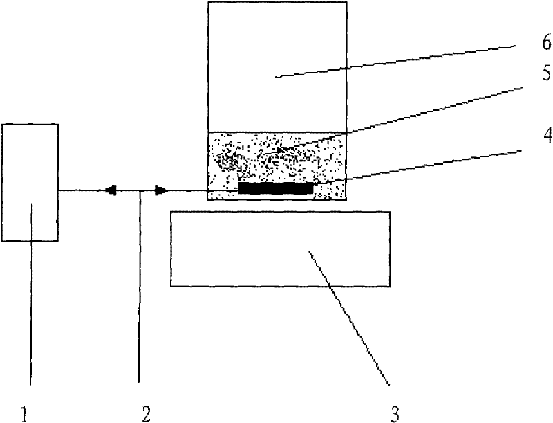
[0005] Below in conjunction with accompanying drawing, the present invention will be further described:

[0006] In the figure, 1. Ultrasonic detector, 2. Ultrasonic wave, 3. Magnetic stirring device (with constant temperature), 4. Stirrer, 5. Whole blood sample, 6. Detection cup. As shown in the figure, after the detector is started, the whole blood sample 5 to be tested is first added to the detection cup 6, and then a metal rod-shaped stirrer 4 is placed in the detection cup 6, and the rotating magnet on the magnetic stirring device 3 drives the stirring The stirrer 4 rotates (or in other forms), and the ultrasonic wave 2 generated by the ultrasonic detector 1 detects the movement state of the stirrer 4, so as to obtain the relevant detection results of the blood coagulation ability.


PUM


Abstract

A rapid detection method for detecting a whole blood coagulation function detects the coagulation function of blood by means of ultrasonically detecting conditions of rotation (movement) of a metal bar in a whole blood specimen. In the method, plasma is not required to be separated, whole blood is directly sampled in a detecting cup (anti-coagulation or coagulation), a metal-bar-shaped stirrer (capable of being wrapped by other materials) is arranged in the detecting cup, and during detection, a rotating magnet is additionally mounted at the bottom of the detecting cup. Before the blood is coagulated, the stirrer rotates along with the rotating magnet, and after the blood is coagulated, the stirrer stops rotating due to resistance. An ultrasonic detector is additionally mounted on a side surface of the detecting cup, and a blood coagulation condition is judged by means of detecting rotation/fixation of the stirrer in the blood.

Description

1. Technical field [0001] The invention belongs to the field of clinical medical examination, and belongs to a detection method of blood coagulation function. 2. Background technology [0002] Rapid detection of blood coagulation ability has very important clinical significance, especially for the diagnosis of acute thrombotic diseases. At present, there are many methods for measuring blood coagulation ability, but most of them use separated plasma for detection. The existing coagulation time measurement of whole blood is performed manually, visually, or through the principle of vibration and feedback to detect the coagulation status in the blood. The error of manual operation is large, and it is difficult to guarantee the quality of inspection. The detection of the latter method is rare in China, the equipment is expensive, and its clinical application is limited, and its detection principle is different from that of the present invention. 3. Contents of the invention ...

Claims


Application Information

Patent Timeline
Patent Type & Authority Applications(China)
IPC IPC(8): G01N33/86G01N29/02
Inventor 徐新曹宁詹小勇陈恒博
Owner SINNOWA MEDICAL SCI & TECH
Why Eureka
Try Eureka