Method for segmenting cardiac nuclear magnetic resonance image

A nuclear magnetic resonance image and heart technology, applied in the field of medical image analysis, can solve the problems of segmented left ventricle epicardium leakage, no good solution proposed, mastoid muscle interference, etc.

Inactive Publication Date: 2012-06-20
BEIJING INSTITUTE OF TECHNOLOGYGY
View PDF3 Cites 12 Cited by
  • Summary
  • Abstract
  • Description
  • Claims
  • Application Information

AI Technical Summary

Problems solved by technology

[0004] The difficulty in segmenting the left ventricle of the heart mainly comes from the following three aspects: First, the gray scale of the image is uneven. This uneven gray scale may be caused by the interference of radio frequency pulses or uneven magnetic field strength during the imaging process, or it may be caused by the high-speed movement of blood hitting the myocardial wall caused by; secondly, the interference of the mastoid muscle. Generally speaking, the part of the mastoid muscle connected to the myocardiu

Method used


Image

  • Method for segmenting cardiac nuclear magnetic resonance image
  • Method for segmenting cardiac nuclear magnetic resonance image
  • Method for segmenting cardiac nuclear magnetic resonance image

Examples


Embodiment Construction

[0080] The present invention will be described in detail below in conjunction with the accompanying drawings and specific embodiments.

[0081] This embodiment specifically realizes the cardiac magnetic resonance image segmentation method proposed by the present invention, including the following steps:

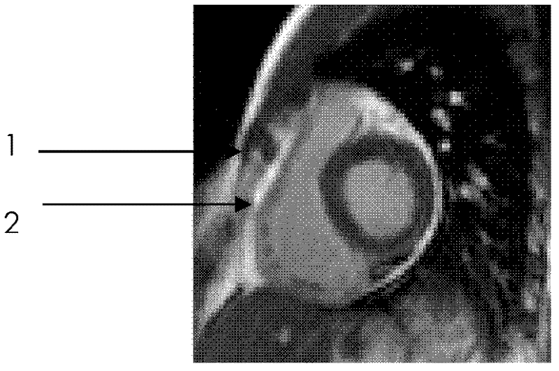
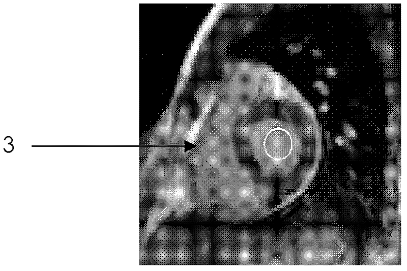
[0082] 1. For the obtained cardiac magnetic resonance images (such as figure 1 Shown) Gaussian filter preprocessing according to the equation Perform Gaussian filter preprocessing on the acquired cardiac MRI images, where I 0 is the input original image structure information, G σ is a two-dimensional Gaussian function with standard deviation σ, Represents a convolution operation. Through Gaussian filter preprocessing, the noise in the image can be effectively filtered, so as to better realize the segmentation of the inner and outer membranes of the left ventricle of the heart.

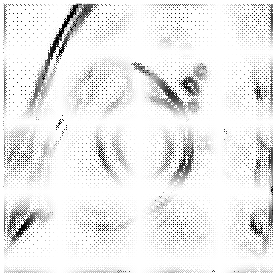
[0083] A cardiac MRI image obtained is attached figure 1 , the preprocessed image is shown...


PUM


Abstract

The invention relates to a method for segmenting a cardiac nuclear magnetic resonance image. The method for segmenting the cardiac nuclear magnetic resonance image comprises the following steps of: 1, performing Gaussian filtering preprocessing on the acquired cardiac nuclear magnetic resonance image; 2, computing an external force field of a generalized gradient vector flow based on expansion neighborhood and noise smoothing on the preprocessed image; 3, defining an initialized outline position of the left ventricular endocardium of the heart; 4, segmenting the left ventricular endocardium of the heart; 5, defining a final segmenting outline result of the left ventricular endocardium of the heart into an initialized outline position of the left ventricular epicardium of the heart; 6, setting the boundary strength of an area surrounded by the endocardium outline in an original boundary graph to be 0, recomputing the external force field of the generalized gradient vector flow based onthe expansion neighborhood and the noise smoothing; and 7, segmenting the left ventricular epicardium of the heart. Based on convolution computation, and by taking energy constraint of an elliptical shape, the method for segmenting the cardiac nuclear magnetic resonance image has the advantages of high computing speed, wide capturing range, strong anti-noise ability, excellent performance in weakboundary protection and deep dented region segmentation, and capability of accurately segmenting the left ventricular endocardium and the left ventricular epicardium of the heart.

Description

technical field [0001] The invention relates to an image segmentation method, in particular to a heart nuclear magnetic resonance image segmentation method, which belongs to the field of medical image analysis. Background technique [0002] Cardiac MRI (magnetic resonance imaging) can provide high-resolution, high-quality images, and accurately describe the anatomical structure and function of the heart. , is of great significance to the early non-invasive diagnosis and accurate prognosis evaluation of cardiovascular diseases. In order to make full use of the anatomical information in the image and provide quantitative and intuitive reference for clinical diagnosis, the intima and adventitia of the left ventricular wall must be segmented first. However, , due to the movement of the heart and the high-speed flow of blood, the image is disturbed by noise, so the segmentation of cardiac MR images is still a problem worthy of further study. [0003] In recent years, the segment...

Claims


Application Information

Patent Timeline
IPC IPC(8): G06T7/00
Inventor 刘利雄赵恒博魏军
Owner BEIJING INSTITUTE OF TECHNOLOGYGY
Why Eureka
Try Eureka