Testing device for identifying antigens and antibodies in biofluids

A technology for biological fluids and testing devices, applied in biological testing, measuring devices, material analysis using immobilized reagents, etc.

Active Publication Date: 2012-07-11
MONASH UNIV
View PDF9 Cites 3 Cited by
  • Summary
  • Abstract
  • Description
  • Claims
  • Application Information

AI Technical Summary

Problems solved by technology

Surprisingly, despite being critical, there is no convenient, low-cost, single-use test that can perform "on the spot" analysis of blood types

Method used


Image

  • Testing device for identifying antigens and antibodies in biofluids
  • Testing device for identifying antigens and antibodies in biofluids
  • Testing device for identifying antigens and antibodies in biofluids

Examples


Embodiment 1

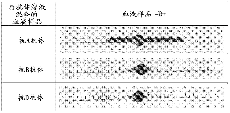
[0045] Example 1: Sequential blood coagulation / coagulation followed by wicking on paper: B+ (two-step method) (see figure 1 )

[0046] Antibodies A and B (Epiclone TM Anti-A, Anti-B and Anti-D; CSL, Australia) solutions. Anti-A and Anti-B are blue and yellow reagents, respectively. "B+" blood was used in this study. Blood samples were filled into plastic bottles with anticoagulant. "B+" blood was separately mixed with pure anti-A and anti-B (used as received) to prepare 100 μL of solution. Paper strips (70mm x 2mm) were made of Whatman #4 filter paper with 2mm unit markings printed on it. The strips were soaked in phosphate buffered saline (PBS). Excess PBS was removed from the strips using standard blotting paper (Drink Coster Blotting, 280GSM). The note was then placed on Reflex Paper (80GSM). Dispense 20 μL of each mixed solution in the center of the paper strip using a graduated micropipette. Photographs were taken after wicking for 4 minutes.

[0047] visible: ...

Embodiment 2

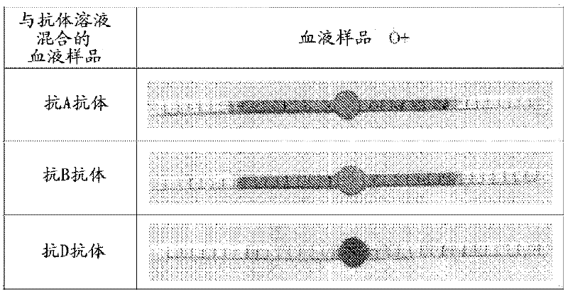
[0054] Example 2: Sequential blood coagulation / coagulation followed by wicking on paper: O+ (two-step method) (see figure 2 )

[0055] Antibodies A and B (Epiclone TM Anti-A, Anti-B and Anti-D; CSL, Australia) solutions. Anti-A and Anti-B are blue and yellow reagents, respectively. "O+" blood was used in this study. Blood samples were filled into plastic bottles with anticoagulant. Mix "O+" blood with anti-A and anti-B separately to prepare 100 μL of solution. Paper strips (70mm x 2mm) were made of Whatman #4 filter paper with 2mm unit markings printed on it. The strips were soaked in phosphate buffered saline (PBS). Excess PBS was removed from the strips using standard blotting paper (Drink Coster Blotting, 280GSM). The note was then placed on Reflex Paper (80GSM). Dispense 20 μL of each mixed solution in the center of the paper strip using a graduated micropipette. Photographs were taken after wicking for 4 minutes.

[0056] visible:

[0057] O+ blood mixed with ...

Embodiment 3

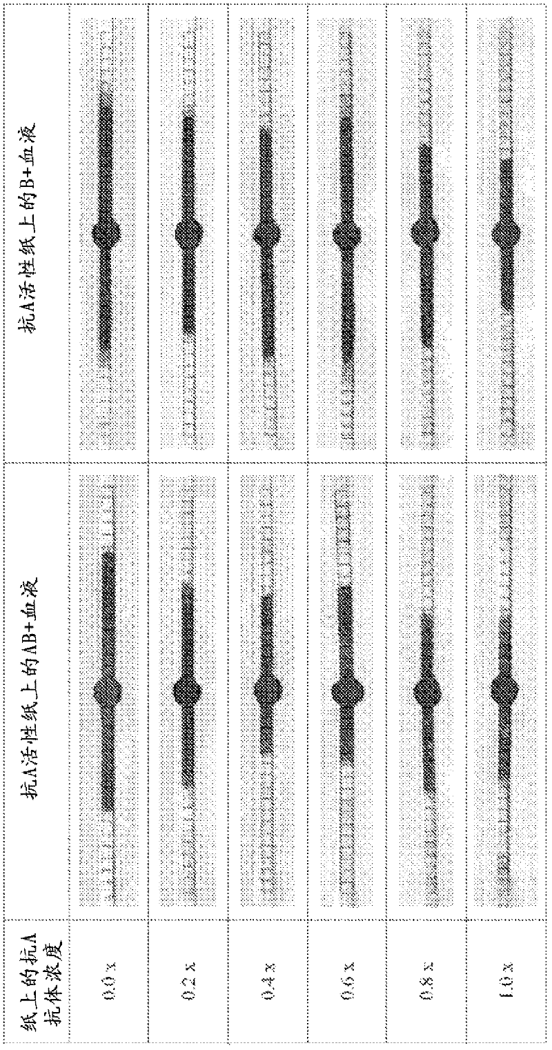
[0063] Example 3: Simultaneous blood coagulation / coagulation followed by paper wicking: effect of antigen concentration (one-step method) (see image 3 )

[0064] In another embodiment of the invention, the paper is first treated with specific antibodies, dried or conditioned prior to exposure to the pure blood sample. This embodiment provides a single step process where the only need is to deposit the blood droplet on the paper. This example also demonstrates the effect of dilute antibody solutions on the wicking and separation properties of blood on paper. Antibody dilution affects the blood (with its antigen) antibody ratio.

[0065] Antibodies A and B (Epiclone TM Anti-A and Anti-B; CSL, Australia) solution. Anti-A and Anti-B are blue and yellow reagents, respectively. "AB+" and "B+" blood were used in this study. Blood samples were filled into plastic bottles with anticoagulant. Paper strips (70mm x 2mm) were made of Whatman #4 filter paper with 2mm unit markings p...


PUM


Abstract

A testing device for identifying an antigen or antibody within a biofluid sample including: a substrate having a hydrophilic surface thereon; the surface including a collection zone, and at least one detection zone extending therefrom; wherein the biofluid sample can be mixed with a specific antigen or antibody, and deposited on the collection zone and transferred by capillary action to the detection zone; the antigen or antibody in the biofluid sample reacting with an appropriate said antibody or antigen thereby resulting in a visual indication within the detection zone.

Description

technical field [0001] The present invention relates to the identification of antigens and antibodies in biological fluids. Although the invention will be described with particular reference to its use in determining human blood typing, it will be appreciated that other applications of the invention are also contemplated. Background technique [0002] Blood is essential to keep tissues alive, and its most important roles are the provision of oxygen and other soluble nutrients, immune protection, and metabolic renewal. Although itself a tissue, blood can be considered chemically as a stable, highly concentrated colloidal suspension consisting of a fluid solution containing many biomolecules (eg, albumin, fatty acids, hormones), metabolites, and electrolytes (serum Red blood cells (red blood cells, 4-6 million / mL, 6-8 μm), white blood cells (leukocytes, 4000-6000 / mL, 10-21 μm), and platelets (150,000-400,000 / mL, 2-5 μm) carried in ) . Some of these biomolecules, such as bin...

Claims


Application Information

Patent Timeline
Patent Type & Authority Applications(China)
IPC IPC(8): G01N33/53D21H25/00G01N33/80A61B5/00G01N30/91
CPCG01N30/90G01N33/80G01N33/558G01N33/54389G01N21/82G01N2021/752G01N2021/757G01N2021/825
Inventor 吉尔·加尼尔沈卫莫希杜什·萨马德·卡恩李煦乔治·图阿斯
Owner MONASH UNIV
Why Eureka
Try Eureka