
Method for processing scalp positioning images of brain tumors

A technology of positioning images and processing methods, applied in image data processing, image analysis, instruments, etc., can solve the problems of inaccurate positioning, lengthening incisions, complicated operations, etc. Effect

Active Publication Date: 2012-07-18
SOUTH CHINA UNIV OF TECH
View PDF4 Cites 21 Cited by
  • Summary
  • Abstract
  • Description
  • Claims
  • Application Information

AI Technical Summary

Problems solved by technology

Because the exact location of the tumor cannot be determined before surgery, a large incision is often required, and sometimes due to inaccurate positioning, the incision may need to be extended during the operation
Stereotactic head frame or neuronavigation system can accurately locate brain tumors, and can correctly guide the direction and depth of surgical approach, but it is expensive and cumbersome to operate, and is not suitable for small and medium hospitals

Method used


Image

  • Method for processing scalp positioning images of brain tumors
  • Method for processing scalp positioning images of brain tumors
  • Method for processing scalp positioning images of brain tumors

Examples


Embodiment

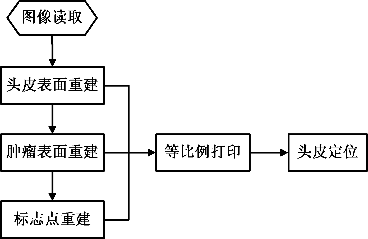
[0035] Such as figure 1 As shown, a brain tumor scalp positioning image processing method of the present invention includes the following steps:

[0036] (1) Preliminarily estimate the scalp projection area of ​​the patient's intracranial tumor, paste two landmarks that can be recognized by the imaging device in the area, obtain a two-dimensional medical image slice set from the imaging device, and reconstruct the three-dimensional contour of the scalp surface. The following steps:

[0037] (1.1) Read the first brain slice image;

[0038] (1.2) Perform mean filtering on brain slice images to remove noise information;

[0039] (1.3) Convert the brain slice image into a binary image;

[0040] (1.4) Calculate the derivatives in the X direction and the Y direction for the binary image, and confirm the pixel points whose derivatives in the X direction and the Y direction are not zero as the contour points of the slice image;

[0041] (1.5) Arrange the contour points sequentially into contou...


PUM


Abstract

The invention discloses a method for processing scalp positioning images of brain tumors, which comprises the following steps of: (1) preliminarily estimating a scalp projected area of a brain tumor of a patient, adhering two mark points which can be identified by an imaging device in the area, obtaining a two-dimensional medical image slice set from the imaging device, and reconstructing a three-dimensional profile of a scalp layer; (2) manually drawing a profile line of the tumor on a two-dimensional image, and reconstructing the surface of the tumor; (3) determining the positions of the two mark points on the two-dimensional image, and reconstructing the mark points; and (4) rotating a profile image of the reconstructed three-dimensional profile image to an appropriate position, and then printing the image in an equal proportion. Compared with the prior art, the method disclosed by the invention is implemented simply; and when the method is applied to the positioning of a brain tumor, the positioning precision meets the demands of tumor resection through a craniotomy, the shortening of operation time and the reduction of surgical traumas are facilitated, and the incision length can be minimized under the premise of guaranteeing good exposures.

Description

Technical field [0001] The invention relates to an image processing method, in particular to a brain tumor scalp positioning image processing method. Background technique [0002] Scalp positioning of brain tumors is an important step before craniotomy, and the accuracy of positioning directly affects the quality of surgery. At present, brain surgeons usually only design craniotomy incisions and remove tumors based on the tumor information shown in CT, MRI and other imaging data, and rely on clinical experience. Because the exact location of the tumor cannot be determined before the operation, a larger incision is often required, and sometimes due to inaccurate positioning, the incision may need to be extended during the operation. Stereotactic headgear or neuronavigation system can accurately locate brain tumors, and can correctly guide the direction and depth of the surgical approach, but it is expensive and cumbersome to operate, and is not suitable for small and medium hospi...

Claims


Application Information

Patent Timeline
IPC IPC(8): G06T7/00G06T7/50G06T17/00
Inventor 杨荣骞何煦佳吴效明白红民
Owner SOUTH CHINA UNIV OF TECH
Learn More