Captive test (CT) image partitioning method based on adaptive learning

A self-adaptive learning, CT image technology, applied in image analysis, image data processing, instruments, etc., to overcome the problem of misclassification, fast segmentation, and improve segmentation effect and efficiency.

Inactive Publication Date: 2012-10-17
SUN YAT SEN UNIV
View PDF2 Cites 24 Cited by
  • Summary
  • Abstract
  • Description
  • Claims
  • Application Information

AI Technical Summary

Problems solved by technology

[0017] The purpose of the present invention is to overcome two problems that CT image segmentation technology exists in the prior art, combine the advantage of the region segmentation method based on classifier mentioned above, propose a kind of CT image segmentation method based on self-adaptive learning, with Adapt to the complex image changes of CT images, so as to solve the above two main problems, and realize accurate and fast segmentation of CT images

Method used


Image

  • Captive test (CT) image partitioning method based on adaptive learning
  • Captive test (CT) image partitioning method based on adaptive learning
  • Captive test (CT) image partitioning method based on adaptive learning

Examples


Embodiment Construction

[0038] specific implementation plan

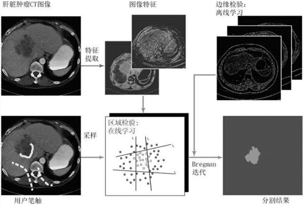
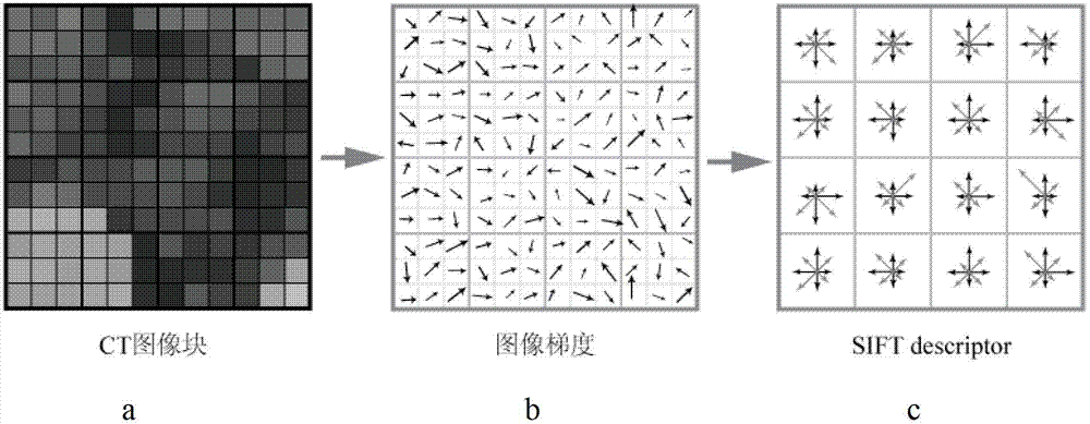
[0039] figure 1It is a system block diagram of the present invention. First, the user marks strokes on the lesion area and non-lesion area on the CT image, and then the system extracts the local grayscale histogram and local SIFT histogram from the image as feature vectors, and uses the pixels on the user's strokes Points are used as training sample points, and the classifier is trained by machine learning method; then the classifier is tested on all pixels on the CT image to obtain the regional item score, and the edge information of the image is iteratively fused using Bregman, and finally the segmentation map of the image is obtained.

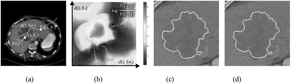
[0040] In the segmentation problem of CT images, the goal is to separate the lesion area displayed in the CT image from other non-lesion areas, that is, to divide the CT image into two areas. Let the area marked with lesion area be the foreground R+, and the area marked with non-lesion area be the back...


PUM


Abstract

The invention discloses a captive test (CT) image partitioning method based on adaptive learning. The CT image partitioning method comprises the following steps of: 1) acquiring a CT image; 2) extracting characteristics of the CT image; 3) inputting strokes for representing a lesion region and a non-lesion region on the CT image by a user; 4) constructing a region model of the image by taking the strokes input by the user as a basis according to the extracted characteristics of the CT image, and constructing an edge model of the image by adopting an edge detection method; and 5) combining the region model and the edge model to construct a new model, calculating the new model to obtain a partitioning result. By adopting the method, a difference between the lesion region and the non-lesion region on the CT image can be effectively described; the method adapts to the complexity of the CT image; the problems caused by low signal-to-noise ratio (high noise) of the CT image are solved; the user can quickly and precisely partition the lesion region on the CT image in an interactive manner with high efficiency; and therefore, the production efficiency of a medical department can be greatly improved.

Description

technical field [0001] The invention relates to the field of medical image segmentation, in particular to the analysis of CT images, the establishment and solution reasoning of CT image segmentation models, online machine learning technology, human-computer interaction technology and the like. technical background [0002] With the advancement of digital imaging technology and image processing technology, the demand for computer-aided diagnosis systems based on digital image processing technology in the medical field is also increasing. The computer-aided diagnosis system based on digital image processing can rely on the current advanced computer processing technology to visualize medical problems and automatically process many complicated things in medicine. It not only provides reliable means for medical staff to make reasonable diagnoses, but also provides protection for patients' lives It also greatly improves the efficiency of medical diagnosis and treatment, making it ...

Claims


Application Information

Patent Timeline
Patent Type & Authority Applications(China)
IPC IPC(8): G06T7/00G06T7/11G06T7/13G06T7/136
Inventor 林倞江波杨巍林梦溪
Owner SUN YAT SEN UNIV
Why Eureka
Try Eureka