Non-partial regularization prior reconstruction method for low-dosage X-ray captive test (CT) image

A CT image, low-dose technology, applied in the field of image reconstruction of medical images, can solve problems such as inability to effectively distinguish edge information from noise, excessive smoothing or step-like effects in reconstructed images, failure to consider prior information of the same patient, etc. , to achieve the effect of improving quality

Active Publication Date: 2012-10-17
SOUTHERN MEDICAL UNIVERSITY
View PDF4 Cites 33 Cited by
  • Summary
  • Abstract
  • Description
  • Claims
  • Application Information

AI Technical Summary

Problems solved by technology

However, the traditional statistical modeling method only introduces the constraints of the local neighborhood of the image itself as prior information, and fails to consider the prior information that can be provided

Method used


Image

  • Non-partial regularization prior reconstruction method for low-dosage X-ray captive test (CT) image
  • Non-partial regularization prior reconstruction method for low-dosage X-ray captive test (CT) image
  • Non-partial regularization prior reconstruction method for low-dosage X-ray captive test (CT) image

Examples


Embodiment Construction

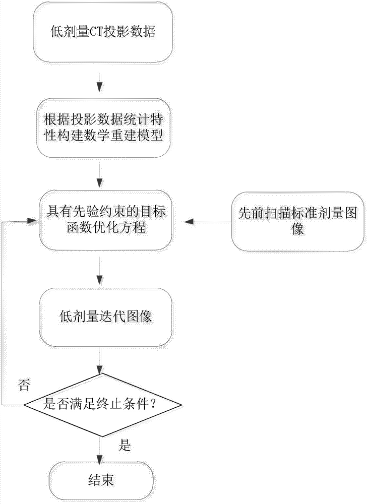
[0040] The non-local regularization prior reconstruction method of a low-dose X-ray CT image of the present invention, its specific implementation steps are as follows figure 1 shown, including the following steps:

[0041] 1. Data acquisition: X-ray CT imaging equipment is used to collect CT medical images scanned by patients previously under standard dose radiation. The medical images under the standard dose radiation are medical images reconstructed by filtered back projection method under clinical standard milliampere-second scanning protocol. Image; use X-ray CT imaging equipment to collect the line integral projection data of the CT medical image of the low-dose radiation of the same patient under the low milliampere-second scanning protocol, and obtain the corresponding correction parameters and system matrix at the same time. Among them, the radiation of the low-dose radiation The dose is 1 / 7 to 1 / 20 of the radiation dose of the standard dose ray, which can be flexibly...


PUM


Abstract

The invention discloses a non-partial regularization prior reconstruction method for a low-dosage X-ray captive test (CT) image. The method comprises the following steps of: (1) acquiring a previously scanned standard dosage image of a patient by X-ray CT imaging equipment; (2) acquiring CT projection data of the patient by the X-ray CT imaging equipment under a Low-mAs scanning protocol, and simultaneously acquiring a corresponding correction parameter and a system matrix; (3) constructing a math model for image reconstruction according to statistical distribution met by the projection data acquired in the step (2); (4) constructing a non-partial regularization prior guided by the previously scanned standard dosage image in the step (1), performing model transformation by adopting a maximum posterior estimation method, and constructing a target function for image reconstruction according to the math model obtained in the step (3); and (5) calculating the target function for CT image reconstruction, which is constructed in the step (4), by adopting an iteration algorithm to finish image reconstruction. By the method, the low-dosage CT image can be reconstructed under the Low-mAs scanning protocol.

Description

technical field [0001] The invention relates to an image reconstruction method of medical images, in particular to a non-local regularized prior reconstruction method of low-dose X-ray CT images. Background technique [0002] The quality of X-ray CT images is closely related to the radiation dose. The higher the radiation dose, the better the image quality. However, excessive X-ray exposure can induce cancer, leukemia or other genetic diseases. Obtaining the best CT imaging diagnostic effect with the least dose is one of the important goals in the field of CT imaging research. [0003] When other scanning parameters remain unchanged, low-dose CT imaging can be achieved by directly reducing X-ray low milliampere-seconds (Low-mAs). However, at this time, a large amount of noise will be introduced into the detection data, which will directly lead to serious degradation of the reconstructed image. , and then affect the clinical diagnosis. A large number of experiments have pro...

Claims


Application Information

Patent Timeline
IPC IPC(8): G06T11/00
Inventor 黄静边兆英张华张蕴婉高杨马建华陈武凡
Owner SOUTHERN MEDICAL UNIVERSITY
Why Eureka
Try Eureka