
Elastography method, elastography system, and biological tissue displacement estimation method and biological tissue displacement estimation system in elastography

A technology of biological tissue displacement and elastic imaging, which is applied in the field of biomedical signal processing, can solve problems such as large amount of calculation, error, and inaccurate displacement estimation, and achieve the effect of improving processing speed and ensuring accuracy

Active Publication Date: 2012-11-07
LEPU MEDICAL TECH (BEIJING) CO LTD
View PDF13 Cites 16 Cited by
  • Summary
  • Abstract
  • Description
  • Claims
  • Application Information

AI Technical Summary

Problems solved by technology

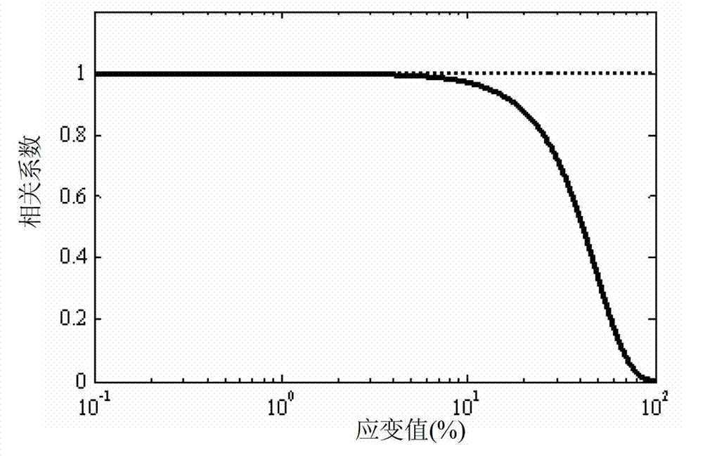
The traditional elastography method is to estimate the displacement of the tissue by calculating the offset of the peak value of the cross-correlation function of the echo signal before and after compression. Due to the axial deformation of the echo signal and other reasons, the de-correlation effect of the signal is generated, which makes the displacement estimation inaccurate and brings a large error. At the same time, some typical applications put forward requirements for real-time signal processing, and the Cross-correlation processing is very computationally intensive

Method used


Image

  • Elastography method, elastography system, and biological tissue displacement estimation method and biological tissue displacement estimation system in elastography
  • Elastography method, elastography system, and biological tissue displacement estimation method and biological tissue displacement estimation system in elastography
  • Elastography method, elastography system, and biological tissue displacement estimation method and biological tissue displacement estimation system in elastography

Examples


Embodiment Construction

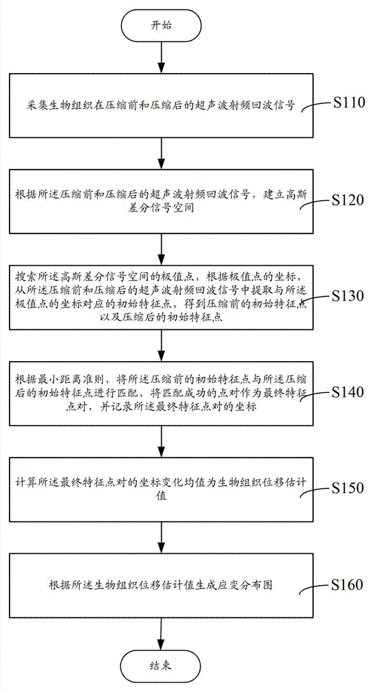
[0048] Such as figure 1 As shown, an elastography method, comprising the following steps:

[0049] S110, collecting ultrasonic radio frequency echo signals of the biological tissue before and after compression;

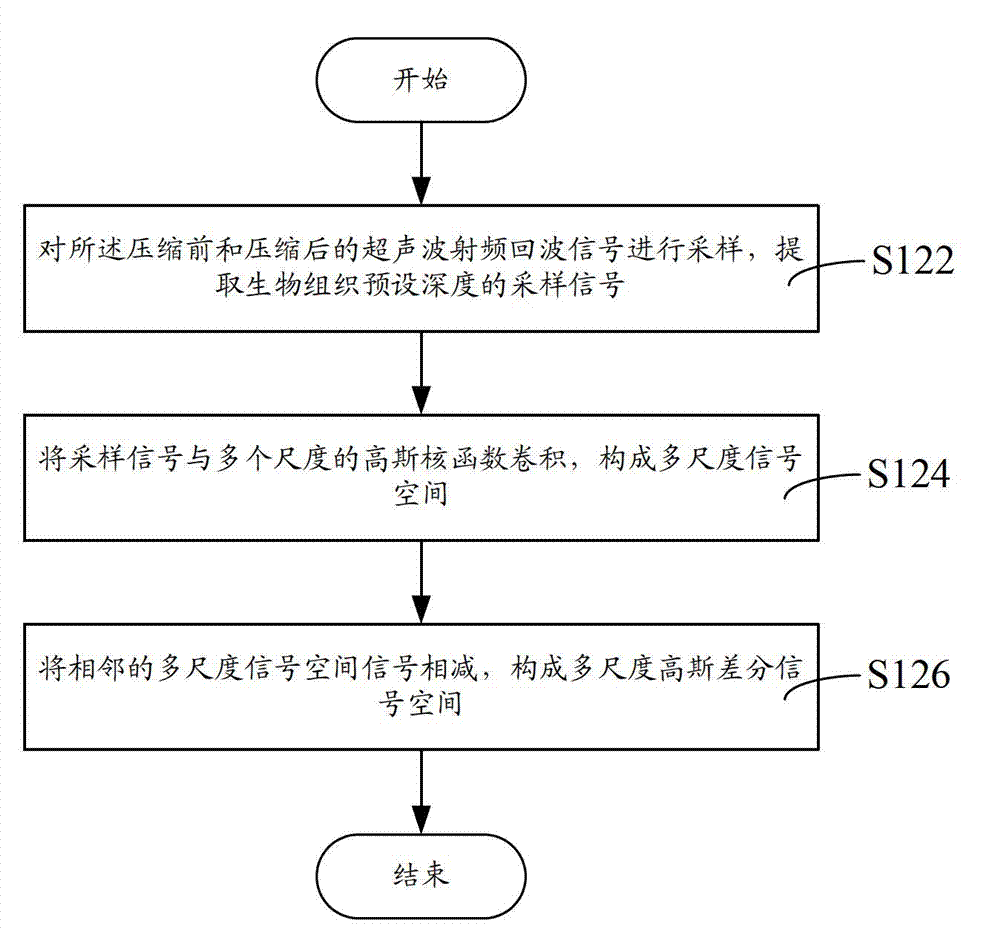
[0050] S120. Establish a Gaussian difference signal space according to the ultrasonic radio frequency echo signals before and after compression;

[0051] S130. Search for the extreme points in the Gaussian difference signal space, and extract initial feature points corresponding to the coordinates of the extreme points from the ultrasonic radio frequency echo signals before and after compression according to the coordinates of the extreme points , get the initial feature points before compression and the initial feature points after compression;

[0052]S140. According to the minimum distance criterion, match the initial feature point before compression with the initial feature point after compression, use the successfully matched point pair as the final feature poi...


PUM


Abstract

The invention discloses an elastography method, an elastography system, and a biological tissue displacement estimation method and a biological tissue displacement estimation system in elastography. The biological tissue displacement estimation method in the elastography comprises the following steps of: acquiring ultrasonic radio frequency echo signals of biological tissues before and after compression; establishing a Gaussian differential signal space according to the ultrasonic radio frequency echo signals before and after compression; searching extreme points of the Gaussian differential signal space, and extracting initial feature points which correspond to coordinates of the extreme points from the ultrasonic radio frequency echo signals before and after compression according to the coordinates of the extreme points so as to obtain initial feature points before the compression and initial feature points after compression; matching the initial feature points before the compression with the initial feature points after compression according to the minimum distance criterion, and recording coordinates of a successfully matched point pair which is taken as a final feature point pair; and calculating a mean value of coordinate transformation of the final feature point pair to obtain a biological tissue displacement estimation value. By the method, the processing accuracy can also be ensured while the processing speed is greatly improved.

Description

technical field [0001] The present invention relates to biomedical signal processing, in particular to an elastic imaging method and system and a biological tissue displacement estimation method and system therein. Background technique [0002] Ultrasound imaging technology has the advantages of no trauma, no ionizing radiation, low cost, and dynamic real-time. It has been widely used in the field of clinical medicine and has become an effective means for early diagnosis of various diseases. The elastic modulus (or hardness) of biological tissue depends on its molecular composition and corresponding microscopic tissue structure, and is closely related to its biological characteristics. There are often differences in elastic modulus (or hardness) between diseased tissue and normal tissue. Ultrasound elastography for the purpose of detecting the mechanical properties of biological tissues is an ultrasound imaging technology that has emerged in recent years. The network signal...

Claims


Application Information

Patent Timeline
IPC IPC(8): A61B8/08
Inventor 肖杨郑海荣孟龙沈洋
Owner LEPU MEDICAL TECH (BEIJING) CO LTD
Learn More