Method for tracking and extracting blood vessels from angiography image full-automatically

A technique of angiography image, extraction method

Active Publication Date: 2012-12-12
BEIJING INSTITUTE OF TECHNOLOGYGY
View PDF4 Cites 26 Cited by
  • Summary
  • Abstract
  • Description
  • Claims
  • Application Information

AI Technical Summary

Problems solved by technology

In general, it is necessary to manually mark on the center line of the blood vessel. When a large number of seed points are required, it is laborious and laborious, and the accuracy of the marking results is not high
[0009] 3. Due to the error between the ridge points that can b

Method used


Image

  • Method for tracking and extracting blood vessels from angiography image full-automatically
  • Method for tracking and extracting blood vessels from angiography image full-automatically
  • Method for tracking and extracting blood vessels from angiography image full-automatically

Examples


Embodiment Construction

[0025] The advantages and spirit of the present invention can be further understood through the following detailed description of the invention and the accompanying drawings.

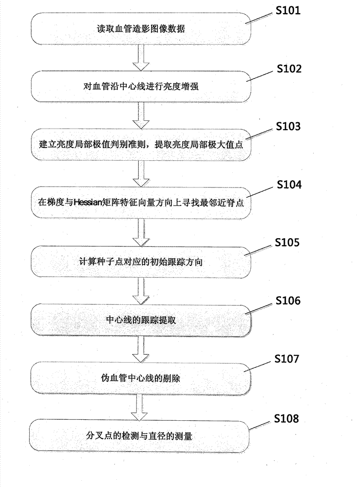
[0026] Step S101, read angiography image data, the data can be a DICOM serial slice image, or a single two-dimensional angiography image.

[0027] Step S102, enhance the contrast image in multi-scale space, wherein the enhancement function at the corresponding scale s is:

[0028]

[0029] Step S103, detecting local maximum points of brightness in the enhanced image, and detecting (x, y) pixel points satisfying the following conditions:

[0030]

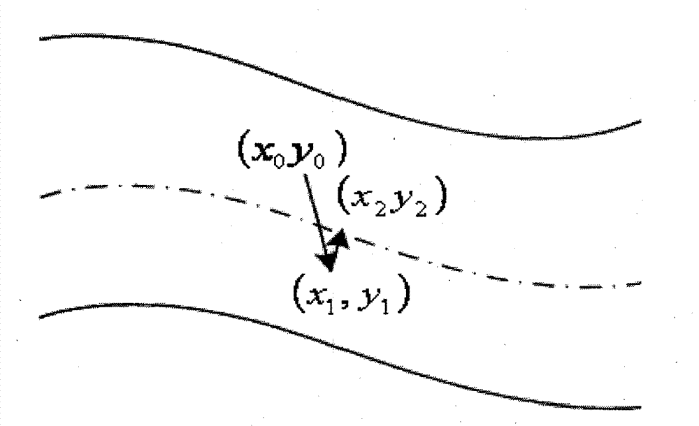
[0031] Step S104, at the obtained brightness local maximum point (x 0 ,y 0 ) normalized gradient (g 1 , g 2 ) in the direction of d 1 Find the brightness maximum point within the distance range (x 1 ,y 1 ),Calculated as follows:

[0032] I ( x 1 , ...


PUM


Abstract

The structures of blood vessels can be segmented from a medical image, and the segmentation is significant for research and diagnosis and treatment of vascular diseases. In a conventional blood vessel extracting method, a large amount of labor force are engaged in the operation and calculated amount of related algorithms is large. The invention provides a method for tracking and extracting blood vessels from an angiography image full-automatically. The method comprises the steps of detecting a large amount of seed points which are located on central lines of blood vessels automatically based on the fact that blood vessels in the image is subjected to multi-scale ridge strengthening and calculating original tracking directions of the blood vessels; acquiring a candidate central line assembly through a method in which ridge points are tracked and extracted on the central lines of the blood vessels and rejecting false blood vessel central lines; and finally, analyzing discrete points on the central lines, and determining which point is a branching point of a blood vessel and calculating blood vessel diameter length corresponding to a ridge point on a central line of the blood vessel. The algorithm is high in operating speed, a topological structure of blood vessels can be extracted accurately without manual intervention, and the method can be used in a clinical diagnosis and treatment process of cardiovascular diseases.

Description

technical field [0001] The invention relates to a method for segmenting blood vessels in angiography images, in particular to a fully automatic method for segmenting blood vessels, which belongs to the field of medical image processing. Background technique [0002] With the improvement of human living standards, cardiovascular and cerebrovascular diseases have become the number one cause of death affecting human health. According to the World Health Organization's recent investigation of the "Top Ten Causes of Death" in humans, the number of deaths from cardiovascular and cerebrovascular diseases in the world has accounted for all deaths every year. 23.6% of the population. Therefore, early quantitative diagnosis and risk assessment of cardiovascular and cerebrovascular diseases play a key role in prolonging human life expectancy and improving human quality of life. [0003] Angiography is a method of showing blood vessels with a contrast medium. In clinical diagnosis and...

Claims


Application Information

Patent Timeline
IPC IPC(8): G06T5/00G06T7/00
Inventor 杨健肖若秀刘越王涌天
Owner BEIJING INSTITUTE OF TECHNOLOGYGY
Why Eureka
Try Eureka