Fundus image vascular segmentation method based on phase congruency

A fundus image and consistency technology, applied in the field of image processing, can solve the problems of inability to correctly segment blood vessels, insensitivity to image brightness and contrast, and achieve the effect of improving the correct segmentation rate of blood vessels, filtering out noise, and good anti-noise performance.

Active Publication Date: 2013-03-20
TIANJIN POLYTECHNIC UNIV
View PDF2 Cites 24 Cited by
  • Summary
  • Abstract
  • Description
  • Claims
  • Application Information

AI Technical Summary

Problems solved by technology

[0004] The purpose of the present invention is to overcome the above-mentioned deficiencies in the prior art, and propose a method for segmenting blood vessels in

Method used


Image

  • Fundus image vascular segmentation method based on phase congruency
  • Fundus image vascular segmentation method based on phase congruency
  • Fundus image vascular segmentation method based on phase congruency

Examples


Example Embodiment

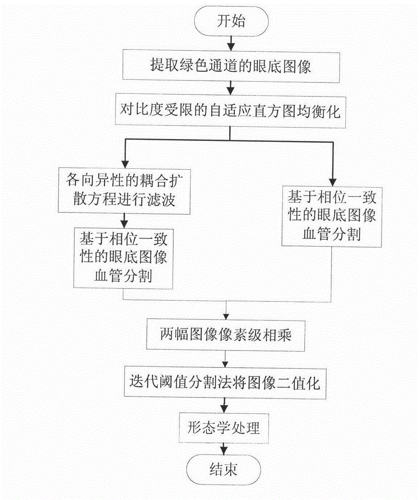
[0030] The flow chart of the present invention is as figure 1 As shown, the green channel of the fundus image is first extracted, and the contrast of the image is enhanced by contrast-limited adaptive histogram equalization (CLAHE); the anisotropic coupled diffusion equation is used to filter to improve the definition of blood vessels; based on phase consistency The algorithm performs blood vessel segmentation on the fundus images with or without the anisotropic coupling diffusion equation filtering; then the pixel-level multiplication of two blood vessel images based on the phase consistency (PC) method; finally, the multiplied image After valueization, the image is optimized with mathematical morphological operations. The specific implementation process of the technical solution of the present invention will be described below in conjunction with the accompanying drawings.

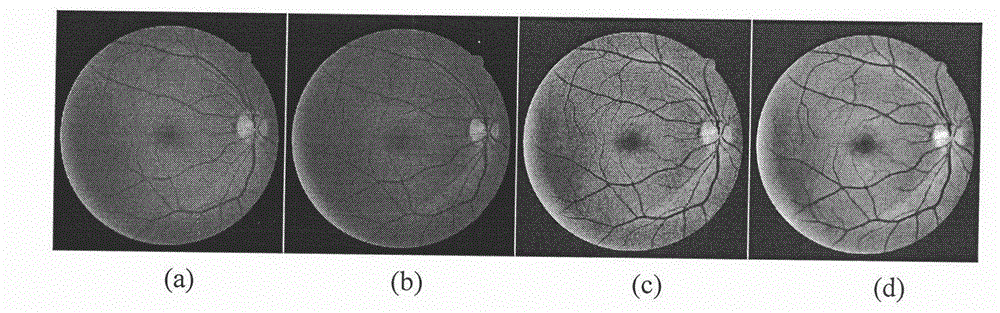
[0031] 1. Extract the green channel of the fundus image;

[0032] Enter as figure 2 The color fun...


PUM


Abstract

The invention discloses a fundus image blood vessel segmentation method based on phase congruency and mainly overcomes the defect that a traditional method can not be used to accurately segment blood vessels in fundus images. The fundus image vascular segmentation method base on the phase congruency can be simultaneously used to segment small blood vessels of most tips. The method comprises the steps: (1) extracting green channels of the fundus images, (2) enhancing the contrast ratio of the images through contrast limited adaptive histogram equalization (CLAHE), (3) filtering the fundus images through the anisotropic coupled diffusion equation, (4) segmenting the blood vessels of the fundus images filtered or not filtered through the anisotropic coupled diffusion equation in a phase congruency algorithm, (5) multiplying pixel-levels of results of vessels, of two fundus images, segmented based on the phase congruency algorithm, (6) processing the images in a binaryzation mode through the iterative threshold segmentation method, (7) optimizing the images in the mathematical morphology method. The fundus image vascular segmentation method has significant application values in fields of three-dimensional splicing of the fundus images and judging existence of diabetes mellitus and severity of diabetes mellitus.

Description

technical field [0001] The invention belongs to the technical field of image processing, and relates to a method for segmenting blood vessels of fundus images based on phase consistency, which can be used for segmenting blood vessels of fundus images with different degrees of normal and pathological changes. Background technique [0002] Changes in the structural features of fundus images can directly reflect the severity and treatment of diabetes mellitus. Blood vessels in fundus images are the most stable and main structures that can be detected in fundus images. When visual diseases occur in eye organs, the diameter, color, and curvature of blood vessels in fundus images will be abnormal. Therefore, the segmentation of blood vessels in fundus images is a very important research problem. Due to hardware limitations, fundus images obviously exhibit the following characteristics: uneven illumination, low contrast between blood vessels and background, and many image noise po...

Claims


Application Information

Patent Timeline
IPC IPC(8): G06T7/00
Inventor 肖志涛耿磊张芳吴骏罗君
Owner TIANJIN POLYTECHNIC UNIV
Why Eureka
Try Eureka